3.5 Respiratory System
Overview
The primary functions of the respiratory system are smell, air conduction, and the exchange of oxygen and carbon dioxide between the animal and the environment, referred to as respiration (Jennings & Premanandan, 2017). It also plays a role in maintaining the acid-base balance. Although cells require oxygen, it is actually the accumulation of carbon dioxide that drives the respiratory system to breathe. This system includes the muscles used to move air in and out of the lungs, the structures involved in the movement of oxygen and carbon dioxide, and the microscopic gas exchange that occurs within the lungs.

Path of Air
Clinical discussion of the respiratory tract is generally divided into the upper respiratory tract and the lower respiratory tract. The upper respiratory tract is composed of the nose, pharynx, and larynx, whereas the lower respiratory tract includes the trachea, bronchi, bronchioles, and alveoli (Jennings & Premanandan, 2017).
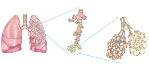
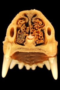
Upon inspiration, air enters the body through the nostrils (nares) into the nasal cavity located inside the nose where the air is warmed to body temperature and humidified by moisture from mucous membranes. These processes help equilibrate the air to the body conditions, reducing any damage that cold, dry air can cause. Particulate matter that is floating in the air is removed in the nasal passages by hairs, mucus, and cilia. From the nasal cavity, air passes through the pharynx (throat) and the larynx (voice box) as it makes its way to the trachea. The main function of the trachea is to funnel the inhaled air to the lungs and the exhaled air back out of the body. The end of the trachea divides into two bronchi that enter the right and left lungs. Air enters the lungs through the primary bronchi. The primary bronchus divides, creating smaller and smaller bronchi called bronchioles as they split and spread through the lung. At the end of each bronchiole are alveolar sacs, each containing multiple alveoli, which is where gas exchange occurs. Oxygen diffuses into the circulatory system from the alveoli, whereas carbon dioxide diffuses from the circulatory system to the lungs. Carbon dioxide is the waste product that cells produce as they use oxygen. Exhaling follows the opposite pathway to inspiration (Fowler et al., 2013).
Take a Breath
Breathing is the inhalation and exhalation of air. Inhalation or inspiration is drawing air in, and exhalation or expiration is releasing the air.

Fun Facts
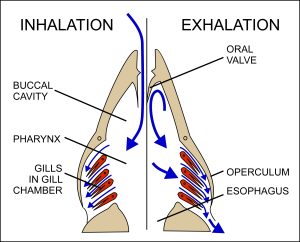
Fish and many other aquatic organisms have evolved gills to take up dissolved oxygen from water. Gills are thin tissue filaments that are highly branched and folded. When water passes over the gills, the dissolved oxygen in water rapidly diffuses across the gills into the bloodstream. The circulatory system can then carry the oxygenated blood to the other parts of the body (Hinic-Frlog, n.d.).

CrashCourse. (2015, August 24). Respiratory system, part 1: Crash Course anatomy & physiology #31 [Video]. YouTube. https://www.youtube.com/watch?v=bHZsvBdUC2I&t=16s
Structures of the Respiratory System
Nose: The main entrance and exit for the respiratory system.

Epiglottis: A very flexible, elastic piece of cartilage that covers the opening of the trachea when swallowing to prevent food and liquid from entering the trachea.
Pharynx: A tube made up of skeletal muscle and lined with mucous membrane that begins in the nasal cavity and ends at the larynx; also known as the throat.
Larynx: Also known as the voice box; a cartilaginous structure that connects the pharynx to the trachea. It plays an important role in air conduction, vocalization, and obstructing the passage of food into the trachea during swallowing via the epiglottis (Jennings & Premanandan, 2017).
Trachea: Also known as the windpipe; extends from the larynx to the lungs and branches at the end into the right and left bronchi. It is made of incomplete rings of cartilage and smooth muscle. The cartilage provides strength and support to the trachea to keep the passage open (Fowler et al., 2013.).
Put a ring on it!
You can feel the rings of cartilage of your own trachea. If you gently palpate the front of your neck/throat region, you can feel a firm tube (your trachea), as well as the rings of cartilage that support it.
Lungs: main organ of respiration allowing for gas exchange.
Bronchi: Air conduction tubes within each lung that branch into smaller tubes called bronchioles. The bronchioles end in alveolar sacs, which contain alveoli.
Alveoli: The air sacs where gas exchange occurs
Diaphragm: A dome-shaped muscle located at the base of the lungs, dividing the thoracic and abdominal cavities. Breathing is dependent on the contraction and relaxation of this muscle.
Vocal cords/vocal folds: are two bands of flexible muscle tissue located inside the larynx that vibrate to make sounds.
| COMBINING FORM | MEANING | EXAMPLES USED IN VETERINARY MEDICINE |
|---|---|---|
| alveol/o | alveoli (air sacs) | alveolar |
| bronch/o, bronchiol/o | bronchial tubes | bronchial |
| cyan/o | blue | cyanotic |
| laryng/o | larynx (voice box) | laryngoscope |
| nas/o, rhin/o | nose | nasal |
| pharyng/o | pharynx | pharyngectomy |
| pneum/o, pneumon/o, pulmon/o | lung | pneumonia |
| trache/o | trachea (windpipe) | tracheostomy |
Common Pathological Conditions of the Respiratory System
Asthma: A chronic disease characterized by inflammation of the airways.
Epistaxis: A nosebleed.
Hemothorax: A collection of blood in the pleural cavity, which is the space between the chest wall and the lung. The most common cause is chest trauma, but hemothorax can also result from abnormal blood clotting or thoracic surgery.
Pleural effusion: An accumulation of fluid in the pleural space.
Pneumonia: An infection involving one or both lungs.
Pneumothorax: An accumulation of air in the space between the pleura, the two-layered membrane that covers the lungs.
Pulmonary edema: Edema within one or both lungs.
Rhinitis: inflammation of the nose.
Stenotic nares: Narrowed nostrils that reduce airflow.

Upper respiratory infection (URI): An infection of the nose, pharynx, and/or larynx
Example
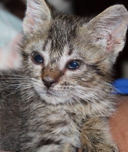
An upper respiratory infection (URI) in kittens can be a potentially life-threatening condition. If left untreated, the kitten runs the risk of struggling to get air into the lungs. Clinical signs can include nasal and ocular discharge, coughing, and dyspnea.

Exercise
What non-infectious respiratory pathology is most often diagnosed in cats?
Answer: Asthma
Common Procedures
Auscultation: The act of listening to the sounds from the heart, lungs and trachea, and other organs, typically with a stethoscope; pulmonary auscultation is listening to the lungs.
Endotracheal intubation: The placement of a flexible plastic tube into the trachea (windpipe) to maintain an open airway; often placed during anesthesia to deliver oxygen and drugs in a gaseous form.
Laryngoscopy: A procedure used to visualize the larynx; often done before placing an endotracheal tube to locate the trachea.
Respiratory rate (RR): The number of breaths an animal takes each minute; an abnormal RR could indicate a respiratory problem.
Respiratory tract radiograph: An image produced on a sensitive plate or film by X-rays, gamma rays, or similar radiation used to examine the respiratory tract.
Thoracocentesis: The aspiration of fluid from the thoracic cavity (Anspaugh et al., 2022).
Tracheostomy: An incision made into the trachea.
Acronyms
CO2: carbon dioxide
CPR: cardiopulmonary resuscitation
ET: endotracheal
O2: oxygen
O2 Sat: oxygen saturation; percentage of oxygen in the bloodstream
RR: respiratory rate (see video below)
URI: upper respiratory infection
shaunablois1. (2020, November 5). Thorax: VETM 3430 [Video]. YouTube. https://www.youtube.com/watch?v=hUVLPGiNxLE&ab_channel=shaunablois1
Additional Respiratory Terms
Anoxia: A lack or loss of oxygen.
Apnea: Not breathing; without breathing.
Antitussive: against cough; medically used to name cough medicine.
Aspiration: To “breathe in” a foreign substance (e.g., when your food “goes down the wrong pipe”).
Bradypnea: A below-normal respiratory rate.
Cyanotic: A bluish or purplish discolouration caused by oxygen deficiency in the blood; the gums will often appear cyanotic if the patient is not breathing well.
Dyspnea: Painful or laboured breathing.
Diaphragmatic: pertaining to the diaphragm.
Nasogastric: pertaining to the nose and the the stomach
Pulse Oximeter (Pulse Ox): an instrument to measure oxygen
Tachypnea: An above-normal respiratory rate.
Exercise
Attribution
Unless otherwise indicated, material on this page has been adapted from the following resource:
Sturdy, L., & Erickson, S. (2022). The language of medical terminology. Open Education Alberta. https://pressbooks.openeducationalberta.ca/medicalterminology/, licensed under CC BY-NC-SA 4.0
References
Anspaugh, K., Goncalves, S., Jackson-Osagie E., & Smith, S. Q. (2022). Medical terminology: An interactive approach. LOUIS: The Louisiana Library Network. https://louis.pressbooks.pub/medicalterminology/, licensed under CC BY 4.0
Fowler, S., Roush R., Wise, J., Reeves, N., DeSaix, J., Kuehner, B., Leady, B., Boggs, L., Broverman, S., Byres, D., Marcus, B., Mhlanga, F., Mignone, M., Nash, E., Newton, M., Oliveras, D., Piperberg, J., Reisenauer, A., Rumfelt, L., Belk, M. … Zoubina, E. (2013). Concepts of biology. OpenStax. https://openstax.org/details/books/concepts-biology, licensed under CC BY 4.0
Hinic-Frlog, S. (n.d.). Introductory animal physiology. University of Toronto Mississauga. https://ecampusontario.pressbooks.pub/introanimalphysiology, licensed under CC BY 4.0
Jennings, R., & Premanandan, C. (2017). Veterinary histology. Ohio State University. https://ohiostate.pressbooks.pub/vethisto, licensed under CC BY-NC 4.0
Image Credits
(images are listed in order of appearance)
202008 lung detailed by DataBase Center for Life Science (DBCLS), CC BY 4.0
20160428 dog border collie panting (cropped) by IketaniDaisei, CC BY-SA 4.0
Breathing in fish by Cruithne9, CC BY-SA 4.0
Dog nose teeth anatomy by katja, Pixabay licence
Kitten with URI by ashalightbearer, Pixabay licence
Bouledogue by JacWehrlin, CC BY-SA 4.0
thin hairs within the nostrils
Pertaining to the alveoli
pertaining to the bronchi or bronchioles
Bluish or purplish discolouration caused by oxygen deficiency in the blood
an instrument used to the examine or view the voice box
pertaining to the nose
Removal of part of the pharynx
Pertaining to the lungs; a serious lung infection caused by a virus or bacteria
An incision into the trachea
fluid accumulation
pertaining to the eye
lack of breath, shortness of breath

