4.7 Nervous System
Overview
The nervous system is very complex and is responsible for controlling much of an animal’s activity, including both voluntary and involuntary functions. After it receives stimuli from the environment, the nervous system creates responses to that information. Animals must be able to sense and respond to the environment in which they live if they are to survive. The nervous system is also responsible for taking sensory input and integrating it with other sensations, memories, emotional states, and learning.
The nervous system can be divided into two main components: the central nervous system and the peripheral nervous system. From there, it is further subdivided by functions and components.
CrashCourse. (2015, February 23). The nervous system, part I: Crash Course anatomy & physiology #8 [Video]. YouTube. https://www.youtube.com/watch?v=qPix_X-9t7E&list=PL8dPuuaLjXtOAKed_MxxWBNaPno5h3Zs8&index=9
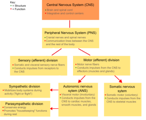
The two main divisions of the nervous system are the central nervous system and the peripheral nervous system.

Central nervous system (CNS): Includes the brain and the spinal cord. The brain controls conscious experiences and regulates homeostasis. The spinal cord controls the sensory and motor pathways.
Peripheral nervous system (PNS): Consists of the cranial and spinal nerves, autonomic nerves, and ganglia. The PNS collects and sends information and has two divisions: the afferent (sensory) division and efferent (motor) division.
- Afferent division: Collects incoming sensory information
- Efferent division: Carries outgoing information and can be further divided into the following systems:
- Somatic nervous system: Responsible for conscious control and for voluntary responses to perception through the use of the skeletal muscles. This allows an animal to respond to their environment with controlled movements.
- Autonomic nervous system: Controls unconscious or involuntary actions, including breathing (lungs), heart rate, smooth muscle, and glands. The two main parts of the autonomic nervous system are the sympathetic nervous system and the parasympathetic nervous system.
- Sympathetic nervous system: Responsible for the “fight-or-flight” response that occurs when an animal encounters a stressful situation. Examples of functions controlled by the sympathetic nervous system include an accelerated heart rate and inhibited digestion. These functions help prepare an animal’s body for the physical strain required to escape a potentially dangerous situation or to fend off a predator.
-
-
- Parasympathetic nervous system: Allows an animal to “rest and digest” and returns the body to a state of homeostasis.
-
Structures
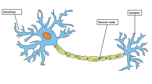
Neurons: Nervous tissue is made up of nerve cells or neurons. They are the basic units of the nervous system and transmit high-speed signals called nerve impulses, much like an electrical current (Hinic-Frlog, n.d.).

- Sensory or afferent neurons: Nerves that carry sensory information from the outside world to the CNS
- Motor or efferent neurons: Nerves that carry signals from the CNS to the muscles and glands
- Associative or interneurons: Nerves that carry nerve impulses from one neuron to another.
The connection or junction between adjacent neurons is called a synapse. The chemical substance that allows this connection is a neurotransmitter, which allows the communication to happen.
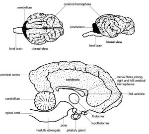
Brain: The main site of nervous control and is highly developed. The major part of the brain lies protected within the skull, also called the cranium. The protective membranes around the brain and spinal cord are called the meninges, and a clear fluid, called cerebrospinal fluid, protects and nourishes the brain tissue (Hinic-Frlog, n.d.).
The brain is divided into three major parts: the cerebrum, cerebellum, and brainstem:
- Cerebrum: The largest part of the brain; coordinates voluntary movement and processes and stores information
- Cerebellum: The second largest part of the brain; coordinates movement
- Brainstem: Regulates the functions of the body that keep the animal alive

Spinal cord: Long nerve tissue that passes through the vertebrae from the brain to the tail. Protective membranes or meninges cover the spinal cord and enclose the cerebral spinal fluid that cushions and nourishes the CNS (Hinic-Frlog, n.d.).
| COMBINING FORM | MEANING | EXAMPLES USED IN VETERINARY MEDICINE |
|---|---|---|
| crani/o | skull | cranium |
| encephal/o | brain | encephalitis |
| myel/o | spinal cord | myelitis |
Common Pathological Conditions of the Nervous System
Ataxia: without coordination
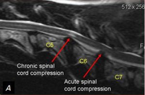
Cervical spondylomyelopathy: Also known as wobbler syndrome; ataxia (poor muscle control) caused by a cervical vertebral malformation that pinches the spinal cord
Example
The abnormal formation of the cervical vertebrae causes ataxia, which often presents as a “wobbly” or swaying gait. This is how cervical spondylomyelopathy got the name wobbler syndrome. Figure 4.32 shows the spinal cord compression (da Costa, n.d.).

Concussion: is a violent shaking of the brain.
Conscious: is a state of being aware or awake.
Encephalitis: Inflammation of the brain
Epilepsy: A neurological disorder characterized by recurrent seizures
Intervertebral disc disease (IVDD): A condition of pain and neurologic deficits resulting from the displacement of part or all of the material in the discs located between the vertebrae
Lethargy: Lack of energy
Meningitis: Inflammation of the meninges, the membranes that cover and protect the brain and spinal cord
Myelitis: Inflammation of the spinal cord
Neuralgia: Nerve pain
Neuropathy: A disease of nerve fibers
Paraplegia: Paralysis in the hind limbs
Paresis: Muscular weakness
Seizure: is sudden involuntary muscle contraction; also called convulsions.
Tetraplegia: Paralysis in all four limbs
Common Procedures
Neurological exam: An exam done in clinic to determine whether an animal has a neurological deficit and to try to determine its source. Some parts of the exam include the following:
- Determining mental status or consciousness; for example, BAR vs. lethargic
- Testing the spinal nerve and cranial nerve reflexes; for example, the patellar reflex, or PLR
- Testing proprioception to determine whether an animal knows where its limbs are located in space
Veterinary Channel. (2023, October 18). How to do a neurological exam on a dog [Video]. YouTube. https://www.youtube.com/watch?v=KTucL_POGl0&t=15s&ab_channel=VeterinaryChannel
Analgesia: Relieving or removing pain
Anesthesia: Using drugs or other substances to cause loss of feeling or awareness
- General anesthesia causes a complete loss of consciousness, typically using either gas inhalation or IV injection of medication.
- Local anesthesia removes sensation in a specific area of the body and is generally injected in the desired area.
- Topical anesthesia removes sensation on the surface of the skin in a specific area and is generally applied to surface of the skin
Disc fenestration: A surgical treatment for IVDD; typically done by a board-certified neurologist in a specialty practice.
Sedation: administration of a medication to decrease response to stimuli.
Acronyms
ANS: autonomic nervous system
BAR: bright, alert, and responsive
CT or CAT scan: computed tomography; diagnostic imaging that shows cross sections of the brain and spinal cord.
CNS: central nervous system
CSF: cerebrospinal fluid
IVDD: intervertebral disc disease
MRI: magnetic resonance imaging
PLR: pupillary light reflex
PNS: peripheral nervous system
QAR: quiet, alert, and responsive
Exercise
Attribution
Unless otherwise indicated, material on this page has been adapted from the following resource:
Sturdy, L., & Erickson, S. (2022). The language of medical terminology. Open Education Alberta. https://pressbooks.openeducationalberta.ca/medicalterminology/, licensed under CC BY-NC-SA 4.0
References
da Costa, R. (n.d.). Wobbler syndrome. Ohio State University, College of Veterinary Medicine. https://vet.osu.edu/research/wobbler-syndrome#:~:text=Wobbler%20syndrome%20is%20a%20neurologic,breeds%20as%20well%20as%20horses
Hinic-Frlog, S. (n.d.). Introductory animal physiology. University of Toronto Mississauga. https://ecampusontario.pressbooks.pub/introanimalphysiology, licensed under CC BY 4.0
Image Credits
(images are listed in order of appearance)
NSdiagram by Fuzzform at English Wikipedia, CC BY-SA 3.0
Example of a neuron by BrunelloN, CC BY-SA 4.0
Anatomy and physiology of animals Longitudinal section through brain of a dog by Sunshineconnelly, CC BY 3.0
Doberman C6-C7 and C5-C6 traction responsive myelopathy A by Filippo Adamo, CC BY-SA 3.0
pertaining to the skull
inflammation of the brain
Inflammation of the spinal cord.
without coordination.

