3.7 Musculoskeletal System
Overview
The musculoskeletal system consists of two systems—the skeletal system and the muscular system—working together to allow movement and providing support to the body. This system includes all the bones, muscles, joints, tendons, and cartilage found in the animal’s body. The purpose of this system is to support the body, allow a wide range of movement, and protect the internal organs.
Some resources show the muscular and skeletal systems as separate; however, for the purposes of this book, they are combined in order to provide a basic overview of their components, functions, and pathologies.
Within the bones is bone marrow that functions to form the red blood cells, white blood cells, and platelets. Muscles contract to allow the animal to move which also results in the production of heat to keep the body warm.
Fish, frogs, reptiles, birds, and mammals are called vertebrates, a name that comes from the bony column of vertebrae (the spine) that supports the body and head. The rest of the skeleton of vertebrates (except fish) also has the same basic design, with a skull that houses and protects the brain and ribs that protect the heart and lungs. All four limbs have a similar bony organization as well (Lawson, 2024).
CrashCourse. (2015b, June 15). Muscles, part 2 – Organismal level: Crash Course anatomy & physiology #22 [Video]. YouTube. https://www.youtube.com/watch?v=I80Xx7pA9hQ&list=PL8dPuuaLjXtOAKed_MxxWBNaPno5h3Zs8&index=23
CrashCourse. (2015a, May 18). The skeletal system: Crash Course anatomy & physiology #19 [Video]. YouTube. https://www.youtube.com/watch?v=rDGqkMHPDqE&list=PL8dPuuaLjXtOAKed_MxxWBNaPno5h3Zs8&index=20
Structures of the Musculoskeletal System
The skeleton is subdivided into two major components:
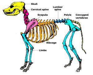
Axial skeleton: The axial skeleton includes all the bones of the skull, vertebrae, sternum, and ribs (bones labelled 1, 2, 4, 5, and 7 in the figure above). It protects the brain, spinal cord, heart, and lungs, and also serves as the attachment site for muscles that move the head, neck, and back.
Appendicular skeleton: The appendicular skeleton includes all the bones of the extremities of the front and hind limbs, plus the bones that attach each limb to the axial skeleton along with the shoulder and pelvic girdle (bones labelled 3, 8, 9, and 10 in the figure above).
Bones
Bones are made of hard, dense connective tissue, and the number of bones varies between species. The purpose of bones is to assist with movement, support the body, protect the organs, and produce blood cells. Bones are in a constant state of building and repairing.
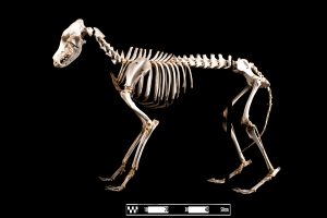
The cranium, also known as the skull, supports the face and protects the brain.
Clinical Insight
The skull has fluid and/or air-filled spaces called sinuses.
The vertebral column consists of a series of bones called vertebrae linked together to form a flexible column, the spine, with the skull at one end and the tail at the other. Each vertebra consists of a ring of bone with spines (spinous processes) protruding dorsally. The spinal cord passes through the hole in the middle, and muscles attach to the spine, making movement of the body possible (Lawson, 2024.)
Paired ribs are attached to each thoracic vertebra (vertebrae associated with the thorax). Each rib is attached ventrally either to the sternum or to the rib in front by cartilage to form the rib cage that protects the heart and lungs. In dogs one pair of ribs is not attached ventrally at all. They are called floating ribs. Birds have large, expanded sternums (called the keel) to which the flight muscles are attached (Lawson, 2024).
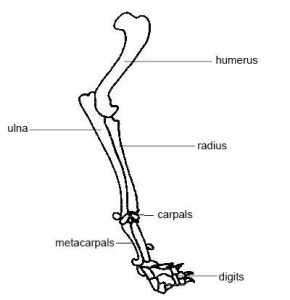
The forelimbs consist of the humerus, radius and ulna, carpals, metacarpals, and the digits or phalanges.
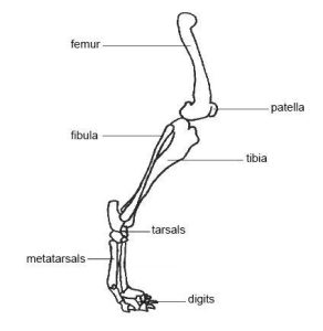
The hind limbs have a similar pattern to the forelimbs. They consist of the femur, tibia and fibula, tarsals, metatarsals, and the digits or phalanges.
Bone Shape and Size
The different bones of the skeleton are divided into groups according to their shape or the way in which they develop.
We have long bones like the femur, radius, and finger bones, as well as short bones like the ones of the wrist and ankle, irregular bones like the vertebrae, and flat bones like the shoulder blade and bones of the skull. Finally, there are bones that develop in tissue separated from the main skeleton, such as the sesamoid bones, which include bones like the patellas (kneecaps) that develop in tendons, and visceral bones that develop in the soft tissue of the penis of dogs and the cow heart (Lawson, 2024).
Muscles
Muscles are one of the four primary tissue types of the body. The body contains three kinds of muscle tissue: skeletal muscle, cardiac muscle, and smooth muscle.
The main function of the muscular system is to assist with movement. Muscles work in pairs; as one muscle contracts, the opposing muscle relaxes. This contraction pulls on the bones and assists with movement. Contraction is the shortening of the muscle fibres, whereas relaxation lengthens the fibres. This sequence of relaxation and contraction is influenced by the nervous system.
Types of Muscle
Skeletal muscles are closely associated with the skeletal system. Also known as striated muscles, they are responsible for voluntary muscle movement, such as running.
Smooth muscles are mainly associated with the walls of internal organs. Also known as visceral muscles, they are responsible for involuntary muscle movement such, as breathing.
Cardiac (heart) muscle, also known as myocardium, is responsible for pumping blood; this pumping creates the heartbeat.

(Molnar & Gair, 2021)
Joints are any place where adjacent bones or bone and cartilage come together to form a connection; also known as articulation.
Cartilage is an elastic connective tissue found at the ends of bones as well as in other locations, such as the tip of the nose.
Tendons are dense, fibrous connective tissues that anchor muscle to bone.
Ligaments are tough, elastic connective tissues that connect bone to bone.
| COMBINING FORM | MEANING | EXAMPLE USED IN VETERINARY MEDICINE |
|---|---|---|
| arthr/o | joint | arthritis |
| cervic/o | neck | cervical |
| crani/o | skull | cranial |
| ligament/o | ligament | ligamentitis |
| muscul/o, my/o | muscle | muscular |
| myel/o | bone marrow | myeloma |
| oste/o | bone | osteoporosis |
| ten/o | tendon | tendonitis |
| vertebr/o | vertebra | vertebral |
Common Pathological Conditions of the Musculoskeletal System
Arthritis: Inflammation of a joint.
Ataxia: A neurological sign consisting of a lack of voluntary coordination of muscle movements.
Dislocation: The displacement of bones in a joint from their normal alignment; also called luxation (Anspaugh et al., 2022).
Fracture: A break in a bone.
Hip dysplasia: a developmental trait characterized by an instability of the hip joint.
Hyperplasia: condition of excessive or increased size of development, formation or growth of cells.
Intervertebral disc disease (IVDD): Refers to several pathological processes involving the discs located between the vertebrae that are used for cushioning; common in dogs, especially in dachshunds.
Osteoarthritis: Inflammation of the bones and joints.
Sprain: Abnormal stretching or tearing of a ligament that supports a joint (Anspaugh et al., 2022).
Strain: Abnormal stretching and tearing of a muscle or tendon (Anspaugh et al., 2022).
Tendonitis: Inflammation of a tendon.
Common Procedures
Amputation: The removal of a limb by trauma, medical illness, or surgery.
Arthrocentesis: A surgical puncture to aspirate fluid from a joint.
Arthroscopy: The visual examination of a joint.
Femoral Head Osteotomy (FMO): A surgical procedure to remove the head of the femur due to pathological conditions such as hip dysplasia or other damages to the hip.
Internal/external fixation: The process of stabilizing an injury (often a fractured bone). This may be accomplished internally (e.g., placing a rod within both parts of the broken bone) or externally (e.g., connecting both parts of the broken bone externally).
Musculoskeletal radiography: An image produced on a sensitive plate or film by X-rays, gamma rays, or similar radiation, and typically used in medical examination of the bones and joints. Radiography is used to identify fractures or tumours, monitor healing, or identify abnormal structures.
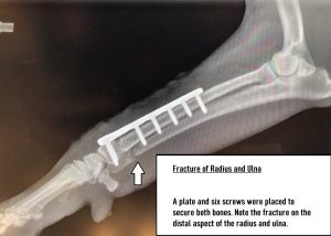
Tibial Plateau Leveling Osteotomy (TPLO): A surgical procedures used to treat cranial cruciate ligament injuries.
Onychectomy: An operation to remove an animal’s claws surgically by means of the amputation of all or part of the distal phalanges, or end bones, of the animal’s toes; also known as declawing.
Acronyms
ACL: anterior cruciate ligament
Fx: fracture, broken bone
DJD: Degenerative joint disease
IVD: Intervertebral disc
IVDD: intervertebral disc disease
MPL: Medial patellar luxation
ROM: range of motion
Additional Musculoskeletal Terms
Ambulatory: ability to walk, relating to walking.
Atrophy: condition of no development.
Brachycephalic: Having a skull that is shorter than average for the species. It is perceived as a cosmetically desirable trait in some domesticated dog and cat breeds, notably the Pug and Persian cats. It can be normal or abnormal in other animal species.
Gait: The way in which the animal walks.
Immobilization: The act of restriction or stopping movement of a limb or bone.
Lameness: limitation or abnormal movement.
Myoma: Tumour of the muscle.
Osteopathy: disease condition of the bones.
Exercise
Attribution
Unless otherwise indicated, material on this page has been adapted from the following resource:
Sturdy, L., & Erickson, S. (2022). The language of medical terminology. Open Education Alberta. https://pressbooks.openeducationalberta.ca/medicalterminology/, licensed under CC BY-NC-SA 4.0
References
Anspaugh K., Goncalves, S., Jackson-Osagie E., & Smith, S. Q. (2022). Medical terminology: An interactive approach. LOUIS: The Louisiana Library Network. https://louis.pressbooks.pub/medicalterminology/, licensed under CC BY 4.0
Lawson, R. (2024). Anatomy and physiology of animals. https://en.wikibooks.org/wiki/Anatomy_and_Physiology_of_Animals, licensed under CC BY-SA 4.0
Molnar C., & Gair, J. (2021). Concepts of biology – 1st Canadian edition. BCcampus. https://opentextbc.ca/biology/, licensed under CC BY 4.0
Image Credits
(images are listed in order of appearance)
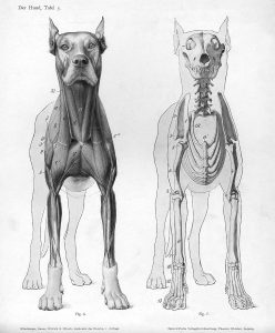
Dog anatomy anterior view by Wilhelm Ellenberger and Hermann Baum, Public domain
Dog skeleton seksjonal by TBjornstad, licensed under CC BY-SA 3.0
Dobermann dog “Canis lupus familiaris” by Museum of Veterinary Anatomy FMVZ USP, edited by Rodrigo.Argenton, licensed under CC BY-SA 4.0
Forelimb dog corrected by Rlawson, licensed under CC BY-SA 3.0
Hind limb dog corrected by Rlawson, licensed under CC BY-SA 3.0
Illu muscle tissues by Arcadian, Public domain
X-ray of forelimb by Kelly Robertson, NorQuest College. Used with permission.
a clotting cell; a blood cell that helps stop bleeding by forming clots; also known as a thrombocyte.
inflammation of the joints
Pertaining to the cervix
pertaining to the skull
Inflammation of a ligament
pertaining to the muscle
A malignant tumor in the bone marrow
is a systemic skeletal disorder characterized by low bone mass
inflammation of the tendon
Pertaining to the vertebra

