4.2 Eyes, Ears, and Additional Special Sense Organs
Overview
In this chapter, we will discuss the senses, including common terminology and abbreviations. The first two major senses are seeing and hearing, which will be discussed in detail. The other senses we will discuss are feeling, smelling, and tasting.
Animals are almost always aware of the stimuli in their environment, meaning the senses are in a constant state of work. Given an animal’s need for survival, their different senses have evolved to adapt to their environment. For example, sharks are electrosensitive, making them able to sense other animals through electromagnetic fields (Molnar & Gair, 2021).
Stimuli such as temperature, pain, pressure, and vibration are called somatosensations. Being aware of orientation and body movement and position are all part of somatosensation (Molnar & Gair, 2021).
The Eye
Vision is the ability to detect light stimuli from the environment and interpret them into images. This is the sense of sight. Within the skull are two bony orbits in which the eyeballs are located. The orbits and the surrounding soft tissue protect the eyes.
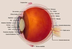
Path of Vision
Light passes through the cornea and enters the eye. The lens refracts the light towards the retina, which is where the receptors are located that convert light to electrical impulses that can be carried via nerves to the brain (Jennings & Premanandan, 2017).
Fun Facts
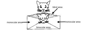
Where the eyes are positioned on an animal’s head can greatly affect their vision. Animals with eyes on the front of their face (commonly predators) have a larger field of binocular vision in front of them, allowing for greater depth perception. This is useful for hunting.
Prey animals tend to have eyes on the sides of their heads, which gives them an overall larger field of view, though with less depth perception. This allows them to detect movement in more directions, which can help them see predators.
CrashCourse. (2015, May 11). Vision: Crash Course anatomy & physiology #18 [Video]. YouTube. https://www.youtube.com/watch?v=o0DYP-u1rNM
Structures of the Eye
Conjunctiva: Epithelial tissue lining the inside of the eyelids and connecting it to the sclera of the eye
Cornea: The front transparent layer of the eye; helps focus light that enters the eye
Eye muscles: Six major muscles are attached to the eye, allowing it to move
Eyelashes: Also called cilia; hairs that protect the eyes from bright light and foreign objects
Eyelids: Upper and lower eyelids cover the eyes during sleep, protect them from foreign objects or too much light, and spread tears over the eye surface
Globe: The entirety of the “eyeball”
Iris: The pigmented ring of smooth muscle that changes the size of the pupil. It constricts the pupil (miosis) in response to bright light and dilates the pupil (mydriasis) in response to dim light.
Lacrimal apparatus (gland or duct): Also called the tear duct; stores, produces, and removes tears
- The nasolacrimal duct drains tears into the nose.
Fun Facts
The pupil varies in shape between species and can be circular (e.g., dog, rabbit), oval (e.g., horse, cow), or vertical (e.g., cat). The colour of the iris can vary with species, age, and sex, and can even differ between eyes or within eyes in the same animal (Jennings & Premanandan, 2017).
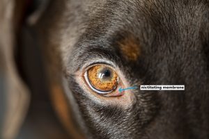
Nictitating membrane: Also called the third eyelid; conjunctiva that is a transparent sheet that moves sideways across the eye from the medial canthus, cleansing and moistening the cornea without shutting out light
Orbit: Deep depressions in the skull that contain the globes
Pupil: The opening at the centre of the eye that allows light to reach the back of the eye
Retina: The layer of photoreceptive and supporting cells on the inner surface of the back of the eye. It is composed of several layers and contains specialized cells for the initial processing of visual stimuli.
Sclera: Visible as the “white” of the eye (Betts et al., 2013); provides support and protection for the eye
| COMBINING FORM | MEANING | EXAMPLES USED IN VETERINARY MEDICINE |
|---|---|---|
| blephar/o | eyelids | blepharitis |
| conjunctiv/o | mucous membrane of the eye | conjunctivitis |
| corne/o | cornea | corneal |
| kerat/o | cornea | keratectomy |
| lacrim/o | tear | lacrimal |
| ocul/o, opt/o, ophthalm/o | eye | ophthalmoscope |
| optic/o | vision | optical |
| retin/o | retina, nervous tissue of the eye | retinal |
| scler/o | white of the eye | scleritis |
(Sturdy, 2022)
Common Pathological Conditions of the Eye
Blepharitis: Inflammation of the eyelid
Cataract: A clouding of the normally clear lens of the eye
Conjunctivitis: Inflammation of the conjunctiva
Corneal ulcer: Injury of the cornea that causes it to become thinner
Ectropion: A medical condition in which the lower eyelid turns outward
Entropion: A medical condition in which the eyelid (usually the lower lid) folds inward
Epiphora: excessive tearing or watering of the eyes; overflow of tears due to decreases drainage
Glaucoma: Increased intraocular pressure (pressure within the globe)
Keratoconjunctivitis sicca: Also called dry eye; a tear disorder that causes the conjunctiva and cornea to dry out
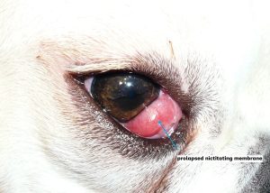
Nictitating gland prolapse: Also called cherry eye; the prolapse of a gland on the medial portion of the eye
Nystagmus: rapid back and forth movement of the eye
Proptosis: A condition in which the globe bulges out of the orbit
Example
Figure 4.4 is an example of cherry eye or ophthalmitis. Repairs to the nictitating membrane could include surgically stabilizing it or surgically removing the membrane itself. This is most common in dog breeds such as the Boston terrier, cocker spaniel, and Pekingese (Hamor, 2023).
Common Eye Procedures and Instruments
Blepharoplasty: Surgical repair of an eyelid
Enucleation: Surgical removal of an eyeball
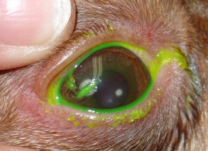
Fluorescein stain: A non-invasive diagnostic procedure used to detect and evaluate corneal integrity (detects corneal ulcers). The bright yellow stain is placed on the cornea, and if there is damage, the dye will stick to it and glow when looked at under a blue light.
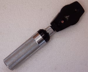
Ophthalmoscope: An instrument used to view the eye
Schirmer tear test: A test used to determine whether the eye produces enough tears to keep it moist
Tonometer: An instrument that measures intraocular pressure
Tonometry: The measurement of intraocular pressure
Eye Acronyms
IOP: intraocular pressure
OD: right eye (“oculus dexter”)
OS: left eye (“oculus sinister”)
OU: both eyes (“oculus uterque”)
STT: Schirmer tear test
Additional Eye Terms
Aqueous humor: liquid found in the eye
Intraocular: Pertaining to within the eye
Lacrimal: Pertaining to the tear duct
Ocular: Pertaining to the eye
Ophthalmologist: A doctor who has special training in diagnosing and treating eye problems
Exercise
The Ear
Hearing, or audition, is the conversion of sound waves into a neural signal that is made possible by the structures of the ear. Audition is important to animals for many different interactions. It enables an animal to detect and receive information about danger, such as an approaching predator, and to participate in communal exchanges like those concerning territories or mating (Hinic-Frlog, n.d.). The ear is also important in maintaining equilibrium or balance.
Auditory Pathway
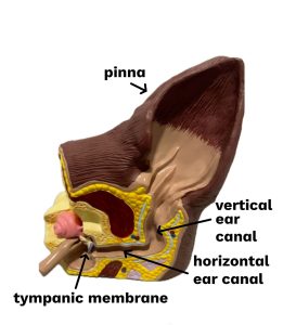
In mammals, sound waves are collected by the external, cartilaginous part of the ear called the pinna (pl. pinnae) or auricle, then travel through the auditory canal and cause vibration of the eardrum (tympanum) (Hinic-Frlog, n.d.). Sound is then conducted via three small bones, which then causes movement of fluid, which is detected via receptors and converted to an electrical signal for the nervous system to transmit.
CrashCourse. (2015, May 4). Hearing & balance: Crash Course anatomy & physiology #17 [Video]. YouTube. https://www.youtube.com/watch?v=Ie2j7GpC4JU
Structures of the Ear
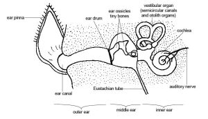
The structure of the ear can be divided into three parts: outer, middle, and inner.
The structure of the ear can be divided into three parts: outer, middle and inner.
Outer ear
The outer ear consists of
- The pinna (auricle), which is the ear flap
- The vertical and horizontal ear canals
Middle ear
The middle ear consists of
- The tympanum (tympanic membrane), which is also known as the eardrum
- The ossicles, which are three small bones that collect and amplify sounds and transfer energy from the moving tympanum to the inner ear
Inner ear
The inner ear is often described as a bony labyrinth because it is composed of a series of canals embedded within the skull that are responsible for hearing and balance.
| COMBINING FORM | MEANING | EXAMPLEs USED IN VETERINARY MEDICINE |
|---|---|---|
| acoust/o
audit/o, aud/i, ot/o |
sound/hearing
ear |
acoustic |
| pinn/i | external ear | pinna |
Common Pathological Conditions of the Ear
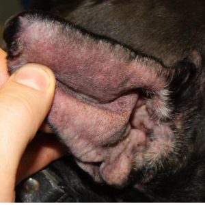
Aural hematoma: A collection of blood within the pinna
Example
An aural hematoma, shown in Figure 4.9, can be caused by trauma or an infection. The clinical signs include a thickened and swollen pinna. A common sign a client will see is the animal shaking its head in an effort to relieve the discomfort. Treatment may include drainage and placing sutures in the ear, as well as a prescription for antibiotics.
Deafness: Complete or partial hearing loss
Otitis externa: An acute or chronic inflammation of the external ear canal
Clinical Insight
Otitis externa is the most common disease of the ear canal in dogs and cats and is much more common in dogs than in cats. It can range from a mild to a severe disease and sometimes extends to the middle ear (Woodward, 2020).
Common Ear Procedures and Instruments
Cytology: The study of cells
Example
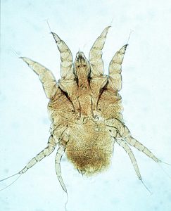
We can swab the external ear canal and then perform cytology to check for ear mites, bacteria, and yeast, all of which can lead to otitis externa.
Otoscope: An instrument used to visually examine within the ear canal
Otoplasty: Surgical repair of the ear
Ear Acronyms
AD: right ear (“auris dextra”)
AS: left ear (“auris sinistra”)
AU: both ears (“aures unitas”)
Additional Ear Terms
Vertigo: condition of the inner ear that causes dizziness.
Vestibular: Pertaining to the vestibule or the sense of balance
Exercise
Additional Special Senses
The first step in sensation is reception, which is the activation of sensory receptors by stimuli from external sources such as being touched or feeling changes in temperature (Hinic-Frlog, n.d.). The senses provide information about the body and its environment. The other senses discussed here are touch and pressure, pain, temperature, proprioception, smell and taste.
Touch and Pressure
Sensory nerves in the skin allow the animal to perceive pressure, temperature, and pain. The nerve endings are found throughout the skin, and some areas are more sensitive than others. Whiskers (vibrissae), for example, are long, tactile hairs that are sensitive to touch and give the animal more information about its environment, such as the size of hole they might want to get into.
Pain
Sensory receptors that respond to pain are known as nociceptors. These receptors are found throughout the entire body. They tell the animal that tissues are dangerously hot, cold, compressed, or stretched or that there is not enough blood flowing in them. The animal may then be able to respond and protect itself from further damage.
Temperature
Nerve endings in the skin that respond to hot and cold stimuli are known as thermoreceptors. The majority of these receptors are in the dermis and will perceive temperatures as hot or cold, allowing the animal to react accordingly.
Proprioception
Proprioception is the sensation that allows animals to know where parts of their body are in space, even if they cannot see them. This is easy to understand when you see an animal walk on all four legs, yet they can’t see their back legs as they walk.
Exercise
Close your eyes and touch your nose. Even without seeing your hand, you are still able to lift it directly to your nose. This is because of proprioception.
Taste
Both taste (gustation) and odour stimuli are molecules taken in from the environment (Hinic-Frlog, n.d). All odours that we perceive are molecules in the air we breathe. Different species have different numbers and types of taste buds, so the same food might taste differently to each species. For example, it’s been determined that cats can’t taste sweet flavours, whereas dogs can.
Smell
Smell, known as olfaction, is a very primitive sense and allows an animal to sense the presence of food, other animals, or chemicals that can impact their survival. Animals also release different smells as messages to communicate between members of their own species and with other species. Many species have a more powerful sense of smell than that of humans. Bears, dogs, and elephants are known for their superior sense of smell.
A pheromone is a chemical released by an animal that affects the behaviour or physiology of other members of the same species. Pheromonal signals can have profound effects on animals that inhale them, but pheromones apparently are not consciously perceived in the same way as other odours. There are several different types of pheromones that are released in urine or as glandular secretions. Certain pheromones are attractants to potential mates, others are repellants to potential competitors of the same sex, and still others play roles in mother-infant attachment. Some pheromones can also influence the timing of puberty, modify reproductive cycles, and even prevent embryonic implantation.
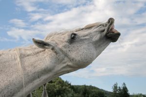
The structure that is involved with the detection of pheromones is the vomeronasal organ. When an animal curls their upper lip, it helps bring the pheromones towards this organ and allows the animal to get a better sense of them (Hinic-Frlog, n.d). This is called the flehmen response and is shown in Figure 4.11.
Exercise
Attribution
Unless otherwise indicated, material on this page has been adapted from the following resource:
Carter, K., & Rutherford, M. (2020). Building a medical terminology foundation. https://ecampusontario.pressbooks.pub/medicalterminology/, licensed under CC BY 4.0
References
Betts, J. G., Young, K. A., Wise, J. A., Johnson, E., Poe, B., Kruse, D. H., Korol, O., Johnson, J. E., Womble, M., & DeSaix, P. (2013). Anatomy and physiology. OpenStax. https://openstax.org/details/books/anatomy-and-physiology, licensed under CC BY 4.0
Hamor, R. E. (2023). The conjunctiva in animals. In Merck veterinary manual: Eye diseases and disorders. Merck. https://www.merckvetmanual.com/eye-diseases-and-disorders/ophthalmology/the-conjunctiva-in-animals
Hinic-Frlog, S. (n.d.). Introductory animal physiology. University of Toronto Mississauga. https://ecampusontario.pressbooks.pub/introanimalphysiology, licensed under CC BY 4.0
Jennings, R., & Premanandan, C. (2017). Veterinary histology. Ohio State University. https://ohiostate.pressbooks.pub/vethisto, licensed under CC BY-NC 4.0
Molnar C., & Gair, J. (2021). Concepts of biology – 1st Canadian edition. BCcampus. https://opentextbc.ca/biology/, licensed under CC BY 4.0
Woodward, M. (2020). Otitis externa in animals. In Merck veterinary manual: Ear disorders. Merck. https://www.merckvetmanual.com/eye-diseases-and-disorders/ophthalmology/the-conjunctiva-in-animals
Image Credits
(images are listed in order of appearance)
Eye with labels by National Eye Institute, Public domain
Anatomy and physiology of animals Well developed binocular vision by Sunshineconnelly, CC BY 3.0
Dog eye head by Sabrinasfotos, Pixabay license
Prolapsed gland of the third eyelid by Joel Mills, CC BY-SA 3.0
Split corneal dystrophy by Joel Mills, CC BY-SA 3.0
Haine Oftamoskop by Janeer, CC BY-SA 3.0
The Ear by Kelly Robertson, NorQuest College. Used with permission.
Anatomy and physiology of animals The ear by Sunshineconnelly, CC BY 3.0
Otitis Othaematom Hund by Kalumet, CC BY-SA 4.0
Psoroptes-cuniculi-ear-canker-mite by Daktaridudu, CC BY-SA 4.0
Flehmendes Pferd 32 c by Waugsberg, CC BY-SA 3.0
sensitive to electromagnetic fields.
Vision that uses both eyes together
pupil constriction
Pupil dilation
medial corner of the eye where upper and lower lids meet
inflammation of the eyelids.
inflammation of the conjunctiva or membrane of the eye.
pertaining to the cornea.
surgical removal of portion of the cornea.
pertaining to the secretion of tears.
an instrument used to examine the eye.
pertaining to vision
pertaining to the nervous tissue of the eye or retina.
inflammation of the white of the eye or sclera.
pressure within the globe
pertaining to sound.
inflammation of the ear
external ear.
pertaining to the ear.
the action or process of receiving something sent, given, or inflicted (oxford dictionary).
Nerve endings that send signals to the central nervous system (CNS) when stimulated
inner layer of the skin.

