3.2 Digestive System
Overview
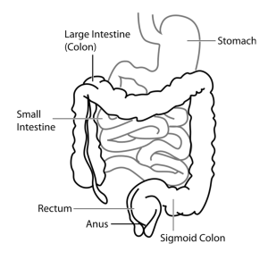
The digestive system, also known as the gastrointestinal (GI) system or alimentary system, is a long, muscular tube that begins at the mouth and ends at the anus. It also has some accessory organs. The functions of the digestive system include the intake, transport, and digestion of food, along with the absorption of nutrients and the removal of physical waste.
Animals can be classified into different groups depending on the type of food they eat. The three groups are herbivores, carnivores, and omnivores.
- Herbivores are animals whose diet is plant-based. Some examples of herbivores include horses, cows, and some bird species.
- Carnivores are animals that eat meat (that is, other animals); these animals include cats, snakes, and sharks.
- Omnivores are animals that eat both plant- and animal-derived foods. Some examples of omnivores are humans, dogs, bears, and chickens.

Path of Digestion
During digestion, food particles are broken down into smaller components both physically (e.g., by chewing) and chemically (e.g., with enzymes). As food is digested, nutrients from the food are absorbed from the digestive tract into the bloodstream.
Food enters the mouth then passes through the pharynx on its way to the esophagus, which transports the food to the stomach using peristalsis for further digestion. Digestion continues in the small intestine where many nutrients are absorbed. The remaining ingesta then passes through the large intestine, and waste is excreted from the body through the anus.
Apart from the main “tube” through which food passes, the digestive system is also comprised of accessory organs that add secretions and enzymes that break down food into nutrients. Accessory organs include the salivary glands, the liver, the pancreas, and the gallbladder.
CrashCourse. (2015, September 7). The digestive system, part 1: Crash Course anatomy & physiology #33 [Video]. YouTube. https://www.youtube.com/watch?v=yIoTRGfcMqM&list=PL8dPuuaLjXtOAKed_MxxWBNaPno5h3Zs8&index=34
Structures of the Digestive System
Mouth (oral/buccal cavity): Begins the process of both mechanical and chemical digestion via mastication, which starts the chemical breaking down of carbohydrates. The mouth includes the lips, cheeks, tongue, gingivae, and teeth.
Cheeks: Composed of muscle and fatty tissue; help keep the food in the mouth.
Gingiva (sing.)/Gingivae (pl.): Also called the gums; consist of mucous membranes that internally line the mouth and surround the teeth
Lips: Form the opening to the mouth. The lips are very tactile, allowing animals to pick up and hold food in their mouth; for example, horses’ lips are sensitive and very mobile for grazing and drinking.
Tongue: Composed of muscle tissue; keeps the food between the teeth during chewing and aids in swallowing. The tongue is also very important for many species for tasting, prehension, licking, vocalizing, and grooming; these functions are more important in some species than others.
The dorsal surface of the tongue has small structures or “bumps” called papillae, which contain the taste buds. A cat uses their papillae to hold saliva to clean their fur, and for lapping up water.
Teeth: Primarily responsible for grinding and breaking down food into small enough pieces to swallow and digest. The number of teeth varies among species.
- Primary or deciduous teeth are the first set of teeth that develop in most species, more commonly known as the “baby teeth.” These teeth usually fall out or resorb into the body at maturity as the permanent “adult” teeth erupt. Retained deciduous teeth are teeth that have not fallen out or been resorbed. In cats and dogs, they are usually extracted at the time of spay or neuter.
- Secondary or permanent teeth are the “adult” teeth.
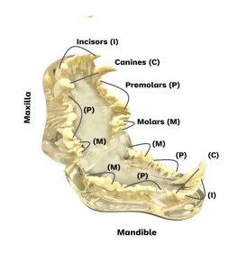
The teeth are arranged in upper (maxilla) and lower (mandible) arcades. There is a dental formula for each species representing the typical distribution of teeth. The four main types of teeth are as follows:
- Incisors: Front teeth used for shearing or cutting
- Canines (“fangs”): Used for tearing and defense
- Premolars: Cheek teeth used for grinding and chewing food
- Molars: Located caudal to the premolars and used for grinding and chewing food

Palate: roof of the mouth. It is divided into two parts:
- Hard palate: boney part under the tissue
- Soft palate: no bone underneath the tissue
Pharynx: Also called the throat; a cavity that is part of both the respiratory and the digestive systems. When food enters the pharynx, involuntary muscle contractions close off the airways to prevent food from entering the lungs. When an animal is not eating, the airways are open for air to enter from the mouth and nose.
Esophagus: A muscular tube that leads from the pharynx to the stomach. When the esophagus reaches the stomach, a ring-like muscle called a sphincter opens and closes to allow the passage of food into the stomach.
Stomach: A sac-like organ that secretes enzymes for chemical digestion. It also has the function of storing and mixing food. Animals with a single stomach, such as humans, dogs, cats, horses, and pigs, are known as monogastric (“one stomach”). Ruminants, such as cows, goats, sheep, camels and giraffes, have multi-chambered stomachs. The most common type of ruminant stomach has four compartments, each with their own function: the reticulum, the rumen, the omasum, and the abomasum. After swallowing, ruminants regurgitate their food to re-chew it. The video below shows a giraffe regurgitating food. Watch as the food is swallowed (moving caudally down the neck), and then regurgitated (moving cranially up the neck).
Fick, J. (April 14, 2013). Giraffe eating cud [Video]. YouTube. https://www.youtube.com/watch?v=c2dzm9u7KTc&ab_channel=Jon%C3%A9Fick
Small intestine: A long, muscular tube that connects the stomach to the large intestine. Further nutrient digestion occurs here, and it is also where most of the nutrient absorption into the bloodstream takes place in the body. It is divided into three parts: the duodenum, jejunum, and ileum (listed from the most proximal to the most distal).
Large intestine: The distal continuation of the small intestine consisting of the cecum, colon, rectum, and anus. The large intestine’s primary functions are the absorption of water and salts, fermentation, and the formation and excretion of feces. The large intestine can vary between species. The terminal end of the large intestine is the rectum, which is connected to the outside world by the anus.
Accessory Organs
Salivary glands: These exocrine glands produce saliva, which is then secreted via ducts into the mouth. The function of saliva is to lubricate food, aid in swallowing, and to start the chemical breakdown of food.
Liver: A large internal organ located caudal to the diaphragm. The liver produces bile, which aids in digestion, removes toxins from the blood, stores some nutrients, and synthesizes proteins.
Clinical Insight
The liver is important in the metabolism of many drugs. Therefore, liver health can affect drug efficacy.
Gallbladder: A sac-like organ located between the lobes of the liver. It stores the bile produced by the liver before it is released into the small intestine.
Fun Fact
Horses have no gallbladder!
Pancreas: Located alongside the small intestine, the pancreas has a mix of exocrine functions, such as secreting digestive enzymes, as well as endocrine functions, such as releasing hormones into the blood.
| COMBINING FORM | MEANING | EXAMPLES USED IN VETERINARY MEDICINE |
|---|---|---|
| abdomin/o | abdomen | abdominal |
| an/o | anus | anal |
| chol/e | bile or gall | cholecystectomy |
| col/o, colon/o | colon | colectomy |
| dent/o, dent/i, odont/o | teeth | dentition |
| enter/o | small intestine | enteritis |
| esophag/o | esophagus | esophageal |
| gastr/o | stomach | gastropexy |
| gingiv/o | gums | gingivitis |
| gloss/o, lingu/o | tongue | glossitis |
| hepat/o | liver | hepatitis |
| labi/o | lip | labial |
| or/o, stomat/o | mouth | oral |
| pancreat/o | pancreas | pancreatic |
| pharyng/o | pharynx | pharyngitis |
| rect/o | rectum | rectal |
(Sturdy & Erickson, 2022)
Common Pathological Conditions of the Digestive System
Ascites: An abnormal collection of fluid in the abdominal cavity.
Bloat: A condition common in ruminants where the rumen distends due to the excessive accumulation of gas produced by the fermentation process. The condition progresses rapidly and can be fatal.
Colic: Is acute abdominal pain. It can be caused by a wide variety of conditions, is often associated with gas and bloating and is potentially life-threatening without treatment in horses.
Coprophagia: The consumption of feces.
Dehydration: A depletion in total body fluids.
Diarrhea: Loose or liquid bowel movements.
Foreign bodies: Non-digestible item(s) that an animal consumes that may not pass through their digestive tract; for example, bones, toys, clothing, or sticks.
Gastric dilatation and volvulus (GDV): An acute, life-threatening emergency most commonly affecting deep-chested large breed dogs. The stomach dilates with gas, then twists and becomes stuck. Commonly affected deep chested breeds including German Shepherds, Great Danes, Irish Wolfhounds, St. Bernards, and Doberman Pinschers.
Gastroenteritis: Inflammation of the stomach and small intestine.
Hemorrhagic Gastroenteritis: acute condition seen in small mammals that causes vomiting and diarrhea. (Hemorrhagic meaning pertaining to the flow or discharge or blood).
Hepatic Lipidosis: is a potentially fatal condition where fat accumulates in the liver; commonly seen in cats after a period of inappetence; Also known as fatty liver disease.
Pica: Eating abnormal objects (ie. cloth, rocks, sticks)
Pancreatitis: Inflammation of the pancreas.
Periodontal disease: An infection of the tissue surrounding the teeth.
Common Procedures
Abdominocentesis: The removal of fluid from the abdominal cavity for testing.
Auscultation: The act of listening to body sounds; for example, for the digestive system, this generally consists of listening to the gut sounds of large animals.
Biopsy: The removal of tissue for examination.
Bloodwork: A lab analysis of blood components.
Drench: Giving liquid medication by forcing the animal to drink it; this term is normally used more often in large animal medicine.
Endoscopy: Using an instrument (an endoscope) to visually examine a hollow body organ or structure; for example, colonoscopy.
Exploratory laparotomy: Exploring or investigating the abdominal cavity and organs via surgery; might be done to find and remove a foreign body that was consumed, to check for masses, or to take biopsy samples.
Fecal testing: A diagnostic tool used to determine abnormalities in stool; for example, parasites.
Fecal Testing Example
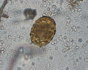
Fecal examinations are a common procedure done in most clinics. Fecal tests are used to detect bacteria, viruses, parasites and abnormal material found in the stool. In Figure 3.3, you can see an intestinal parasite egg found in a fecal floatation. More information can be found in 5.4 Laboratory Terminology.

Float: An instrument used to file or rasp overgrown equine teeth; for more information see 8.5 Large Animal Procedures.
Laparotomy: Cutting into the abdomen.
Nasogastric intubation: Placement of a tube through the nose into the stomach. This is often done in horses who are colicking.
Radiography: An imaging technique used most commonly to visualize internal structures such as bones and organs.
Ultrasound: An imaging technique to visualize of internal body structures that uses high-frequency sound waves to produce an image.
Acronyms
BM: bowel movement
GDV: gastric dilatation volvulus
GIT: gastrointestinal tract
IBD: inflammatory bowel disease
MM: mucous membranes
NPO: nothing by mouth
PD: polydipsia
PO: by mouth
V & D: vomiting & diarrhea (Note: V/D means ventral/dorsal)
Additional Digestion Terms
Absorption: The process of taking digested nutrients into the circulatory system.
Anal Sacs/Glands: Two glands found near the anus of dogs and cats.
Buccal: Relating to the cheek.
Defecation: the process of expelling feces.
Digestion: The process of breaking down food so the body can use the nutrients.
Emesis: To vomit.
Feces: Solid or semisolid waste excreted from the digestive system.
Halitosis: bad breath
Inappetence/anorexic: Having a decreased appetite.
Ingestion: The process of taking food into the body.
Jaundice: Is a condition characterized by yellowing of the mucous membranes and skin due to excessive bilirubin in the body.
Lethargy: A state of tiredness, sleepiness, weariness, fatigue, sluggishness, or lack of energy.
Melena: Dark, tarry stool due to digested blood
Metabolism: the process of changing food into energy for use in the body.
Nausea: Feeling the urge to vomit due to upset stomach.
Obese: Excessive fat accumulation in the body.
Oral: Pertaining to the mouth.
Polyphagia: Increased eating.
Sublingual: Pertaining to under the tongue.
Vomiting: The forceful expulsion of stomach contents.
Clinical Insight
Anal Sacs/Glands are used for scent marking in dogs and cats. They are normally expressed or emptied on their own, but can become impacted or infected and may need to be expressed manually by a DVM or RVT.
Exercise
Attribution
Unless otherwise indicated, material on this page has been adapted from the following resource:
Molnar C., & Gair, J. (2021). Concepts of biology – 1st Canadian edition. BCcampus. https://opentextbc.ca/biology/, licensed under CC BY 4.0
References
Sturdy, L., & Erickson, S. (2022). The language of medical terminology. Open Education Alberta. https://pressbooks.openeducationalberta.ca/medicalterminology/, licensed under CC BY-NC-SA
Images
(images are listed in order of appearance)
Digestive system by Clker-Free-Vector-Images, Pixabay licence
Canine Teeth by Kelly Robertson, NorQuest College. Used with permission.
Ascaris suum egg by Strongyle, CC BY-SA 3.0
is a type of wave-like involuntary muscle movement that occurs in the digestive tract allowing the movement of food and liquid.
material taken into the body via the digestive tract
the process of chewing food
the act of grasping food
pertaining to the abdomen
Pertaining to the anus
surgical removal of the gallbladder
removal of part of the colon
refers to the teeth as a whole
inflammation of the intestines
pertaining to the esophagus
surgical stabilize the stomach to the abdominal wall
inflammation of the gums
inflammation of the tongue
Inflammation of the liver
pertaining to the lips
pertaining to the mouth
Pertaining to the pancreas
Inflammation of the pharynx
pertaining to the rectum
When a horse is experiencing colic (abdominal pain)

