3.4 Cardiovascular System
Overview
The cardiovascular system uses blood to deliver nutrients, oxygen, and hormones to the trillions of cells in the animal’s body and transport waste away from them. The heart, which is the primary organ in this system, pumps blood throughout the body via a network of blood vessels. These three components—blood, blood vessels, and the heart—make up this complex system. Cardiovascular means “pertaining to the heart and blood vessels.”
Path of the Cardiovascular System
Cells use oxygen and release carbon dioxide as a waste product, so the blood leaving them is considered “oxygen-poor”. This oxygen-poor blood from throughout the body enters the right side of the heart via the veins and is pumped to the lungs to be oxygenated. The oxygen-rich blood then returns to the left side of the heart and is pumped out to the rest of body. The cycle then repeats.
Going with the Flow
Here’s a visual to help you understand how blood flows through the body. The blue text represents the blood with low levels of oxygen and high levels of carbon dioxide, while the red text represents blood with high levels of oxygen and low levels of carbon dioxide. The purple font represents that gas exchange happens in that structure. Gas exchange is further discussed in the respiratory chapter.
Organ ⇒ Veins ⇒ Right atrium ⇒ Right ventricle ⇒ Lungs ⇒ Left atrium ⇒ Left ventricle ⇒ Aorta ⇒ Rest of the body
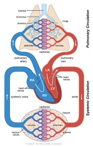
(Fowler et al., 2013)
CrashCourse. (2015, July 6). The heart, part 1 – Under pressure: Crash Course anatomy & physiology #25 [Video]. YouTube. https://www.youtube.com/watch?v=X9ZZ6tcxArI&list=PL8dPuuaLjXtOAKed_MxxWBNaPno5h3Zs8&index=26
Structures of the Cardiovascular System
Heart: A hollow muscular organ that lies between the lungs in the space referred to as the mediastinum. The heart (and lungs) are located in the thoracic/chest cavity. The heart is a pump that provides power to move blood through the body. Electrical currents through the heart are responsible for the contractions that make the heart beat and pump blood.
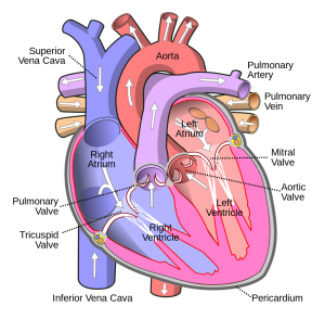
Most mammals have four heart chambers, two on the right side and two on the left side. The cranial chambers are called atria (the singular is atrium) and the caudal chambers are called ventricles (Fowler et al., 2013).
Valves separate the chambers of the heart from each other and from major blood vessels. The atrioventricular (AV) valves are flaps of connective tissue between the atrium and the ventricle, while the semilunar valves are those between the ventricles and the connected major blood vessel (Molnar & Gair, 2021).
The heart pumps blood through the body in a repeating sequence called the cardiac cycle. The cardiac cycle is the flow of blood through the heart coordinated by electrochemical signals that cause the heart muscle to contract and relax. In each cardiac cycle, a sequence of contractions pushes out the blood, pumping it through the body; this is followed by a relaxation phase during which the heart fills with blood. These two phases are called systole (contraction) and diastole (relaxation), respectively (Fowler et al., 2013).
Clinical Insight
If you listen to the heart with a stethoscope, you should hear two sounds, often described as lub-dub. Each of these sounds is actually the valves of the heart closing during the cardiac cycle—the lub is the AV valves closing, and the dub is the semilunar valves closing (Fowler et al., 2013).
Different species have different heart rates. Generally, the smaller and younger the animal, the faster the heart rate.
Follow this link to hear heart sounds as though you were listening through a stethoscope: Cardiac Library
- Listen to both a horse (Normal Equine S3 Sounds) and a cat (Normal 6-year-old Domestic Shorthair Cat). Do you hear the difference in the heart rates?
- Listen to some examples of heart murmurs to hear the “whoosh” between heart sounds indicating blood is flowing through the valve when it shouldn’t be.
Myocardium: The middle and thickest layer of muscle of the heart (the prefix myo- means “muscle”).
Pericardium: The double-walled membrane surrounding the heart (the prefix peri- means “around”).
Blood: Composed of many different compounds, blood can be analyzed using lab tests to determine the various levels of these compounds present in the body.
The primary function of blood is to deliver oxygen and nutrients to the body’s cells and to remove wastes from those same cells. Blood also has other functions, including defence, distribution of heat, and maintenance of homeostasis. For more information, see 4.3 Hematologic System.
Blood vessels: Blood vessels transport blood throughout the body. An artery is a blood vessel that carries blood away from the heart. It branches into smaller vessels called arterioles, and they further branch into tiny capillaries, where nutrients, gases, and wastes are exchanged. The capillaries then combine to form venules, which are small blood vessels that carry blood to a vein, a larger blood vessel that returns blood to the heart.
Blood Vessels
Arteries are blood vessels that move blood away from the heart, while veins move blood towards the heart from other organs. Most arteries carry oxygenated blood to the other organs of the body, while the veins carry deoxygenated blood from the organs back to the heart. One exception to this are the pulmonary arteries and veins. This is because the pulmonary arteries bring deoxygenated blood from the heart to the lungs for gas exchange, while the pulmonary veins are the ones that carry oxygenated blood back to the heart (Figure 3.10 above illustrates this).
RBVH Temp. (2014, March 5). How to check your pet’s vital signs [Video]. YouTube. https://www.youtube.com/watch?v=Lv63ZUTp_IM&t=143s
| COMBINING FORM | MEANING | EXAMPLES USED IN VETERINARY MEDICINE |
|---|---|---|
| cardi/o, coron/o | heart | cardiomegaly |
| hem/o, hemat/o | blood | hematoma |
| phleb/o | vein | phlebotomy |
| vas/o | blood vessel | vasoconstriction |
| ven/o | vein | intravenous |
Common Pathological Conditions of the Cardiovascular System
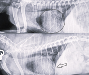
Cardiomegaly: Enlargement of the heart.
Example
Figure 3.9 shows an X-ray of a dog that has been diagnosed with cardiomegaly. Both radiographs show the heart from a right lateral view. The darker area on the X-rays is the lungs and the circled area is the heart. Both the heart and lungs appear abnormal in this X-ray.
Cardiomyopathy: Disease of the myocardium.
Carditis: Inflammation of the heart.
Congestive heart failure: The inability of the heart to pump enough blood through the body.
Cyanosis: condition of blue; tissue and mucous membranes turn blue due to the lack of oxygen.
Edema: The swelling of tissues caused by excess interstitial fluid (Anspaugh et al., 2022).
Heart murmur: An abnormal heart sound associated with blood passing through a valve that should be closed; sounds like a “whoosh” between heart sounds.
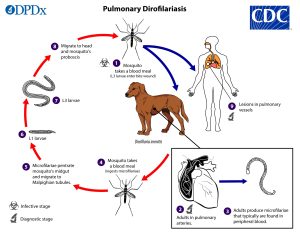
Heartworm: A parasitic worm (Dirofilaria immitis) whose larvae are transmitted by mosquitoes. The worms can affect dogs, cats, and ferrets. They obstruct blood flow through the heart and blood vessels (Root Kustritz, 2022).
Hematoma: A collection of blood outside the blood vessels, usually because of trauma, disease, or surgery (Anspaugh et al., 2022).
Hypoxemia: A lower-than-normal amount of oxygen in the blood.
Ischemia: Lack of blood flow to an area.
Shock: The inability of the body to get oxygen to the tissues and insufficient return of blood to the heart.
Common Procedures
Blood pressure measurement: A measure of the pressure the blood applies to the arteries. Measured both in systole and diastole.
Blood transfusion: Whole blood or blood components given intravenously to the patient.
Capnograph: Instrument used to record carbon dioxide (CO2).
Cardiopulmonary resuscitation (CPR): An emergency technique used if the heart of a patient stops. Applying pressure on the chest can manually compress the blood within the heart into circulation, and respiration is also supported.
Cardiovascular radiograph: An image produced on a sensitive plate or film by X-rays, gamma rays, or similar radiation; used to examine the heart and structures around the heart.
Echocardiography: An ultrasound of the heart; often shortened to “echo” in clinic.
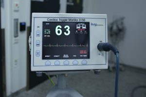
Electrocardiogram (ECG, EKG): A visual record of the electrical impulses in the heart.
Clinical Insight
The electrical impulses in the heart produce electrical currents that flow through the body and can be measured on the skin using electrodes. This information can be observed as an electrocardiogram (ECG, EKG), a recording of the electrical impulses of the cardiac muscle. The ECG can illustrate the rhythm of the heartbeats (Fowler et al., 2013).
Hemostasis: The process of stopping bleeding.
Clinical Insight
Surgeries to do with the cardiovascular system are normally not done in general practice and the patients are sent to specialty clinics with veterinary cardiologists.
Acronyms
BP: blood pressure
BPM: beats per minute
CHF: congestive heart failure
CPR: cardiopulmonary resuscitation
CPA: cardiopulmonary arrest
CV: cardiovascular
ECG or EKG: electrocardiogram or electrocardiograph
HR: heart rate
MM: mucous membranes
Additional Cardiovascular Terms
Arrhythmia: An abnormal rhythm of heart beats. It can also be any form of fibrillation, which is an uncoordinated beating of the heart. An electrocardiogram provides a record of the electrical activity of the heart and can help diagnose this condition.
Blood pressure: The force exerted by the blood against the wall of a vessel.
Bradycardia: An abnormally slow heart rate.
Cardiac auscultation: Listening to a patient’s heart sounds.
Congenital Heart Disease (CHD): is a heart disorder present at birth.
Diastolic blood pressure: The lower value of blood pressure measured during ventricular relaxation.
Hypercapnia: condition of increased or excessive carbon dioxide levels.
Hypocapnia: condition of decreased or deficient carbon dioxide levels.
Hypertension: Abnormally high blood pressure.
Hypotension: Abnormally low blood pressure.
Hypoxia: condition or decreases or deficient levels of oxygen.
Intravenous: Within or into a vein.
Occlusion: process of blockage or closing of a blood vessel or organ.
Pulse: The palpable alternating expansion and recoil of an artery as blood moves through the vessel; an indicator of heart rate.
Pulse oximetry: The measurement of the oxygen level of the blood (oxygen saturation).
Stethoscope: A medical device used for auscultation or listening to the internal sounds of an animal’s body.
Sphygmomanometer: An instrument used to measure blood pressure.
Systolic blood pressure: The maximum value of blood pressure measured following a ventricular contraction.
Tachycardia: An abnormally fast heart rate.
Thrombus: A blood clot.
Vasodilation: condition of widening of the blood vessels.
Exercise
Attribution
Unless otherwise indicated, material on this page has been adapted from the following resource:
Sturdy, L., & Erickson, S. (2022). The language of medical terminology. Open Education Alberta. https://pressbooks.openeducationalberta.ca/medicalterminology/, licensed under CC BY-NC-SA 4.0
References
Anspaugh, K., Goncalves, S., Jackson-Osagie E., & Smith, S. Q. (2022). Medical terminology: An interactive approach. LOUIS: The Louisiana Library Network. https://louis.pressbooks.pub/medicalterminology/, licensed under CC BY 4.0
Fowler, S., Roush R., Wise, J., Reeves, N., DeSaix, J., Kuehner, B., Leady, B., Boggs, L., Broverman, S., Byres, D., Marcus, B., Mhlanga, F., Mignone, M., Nash, E., Newton, M., Oliveras, D., Piperberg, J., Reisenauer, A., Rumfelt, L., Belk, M. … Zoubina, E. (2013). Concepts of biology. OpenStax. https://openstax.org/details/books/concepts-biology, licensed under CC BY 4.0
Molnar C., & Gair, J. (2021). Concepts of biology – 1st Canadian edition. BCcampus. https://opentextbc.ca/biology/, licensed under CC BY 4.0
Root Kustritz, M. (2022). Veterinary preventative medicine. University of Minnesota. https://pressbooks.umn.edu/vetprevmed/, licensed under CC BY-NC 4.0
VetMedux. (n.d.). Cardiac library. CliniciansBrief. https://www.cliniciansbrief.com/custom/cardiac-library
Image Credits
(images are listed in order of appearance)
Blood circulation by Ohio State University, CC BY-NC 4.0
Diagram of the human heart (cropped) by Wapcaplet, CC BY-SA 3.0
Cardiomegaly by Kelly Robertson, NorQuest College. Used with permission.
Diro Pulmonary LifeCycle lg by CDC DPDx, Public Domain
ECG machine by OsloMetX, Pixabay licence
Small veins
blood with oxygen
blood without oxygen
enlargement of the heart
a mass of blood
an incision into a vein
Narrowing of a blood vessel
Pertaining to within a vein
an image produced on a sensitive plate or film by X-rays, gamma rays, or similar radiation, and typically used in medical examination. (wikipedia).
is the tissue fluid that travels between the spaces between the cells of a tissue or an organ
obtaining by applying pressure to the mucous membranes to blanch them, then determining the time it takes to return to normal colour

