8.3 Large Animal Anatomy
Overview
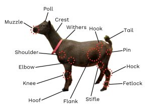
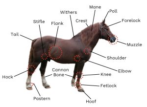
Anatomical terms in large animal medicine are very important to help describe the location of injuries, so it is crucial to have a general knowledge of these topographical markers. Many of the terms are used across large animal species, but a few are more commonly used in some species than others.
The general layout of the skeleton of large animals is very similar to the human skeleton. The table below lists anatomical terms for large animals, along with their human counterpart. One of the main differences between ungulates and humans is the number of toes and fingers. Horses stand on the equivalent of our middle finger while cows, sheep, goats, and pigs stand on the equivalent of two fingers.
From Head to Toe
The following table discusses some of the common anatomy landmarks you will hear in large animal medicine and some of the species they are commonly used for.
If “all” is used in the “Common Species” column, it means this term is used in cattle, horses, sheep, goats, and pigs.
| Anatomical Term | Common Species | Definition |
|---|---|---|
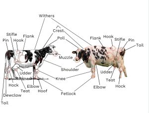
| Muzzle | Horses, cows, sheep, goats | The most rostral portion of the head; the nose and lips |
| Snout | Pigs | The nose |
| Poll | Horses, cows, sheep, goats | Most dorsal aspect of the head |
| Forelock | Horses | Portion of the mane that extends over the face |
| Mane | Horses | Longer, coarser hair growing from the dorsal aspect of the neck |
| Crest | Horses, cows, sheep, goats | Dorsal aspect of the neck running from the skull to the withers |
| Withers | Horses, cows, sheep, goats | Highest point of the back, where the neck meets the back |
| Shoulder | All | Equivalent to human shoulder
Joint where the scapula (shoulder blade) meets the humerus (front limb) |
| Elbow | All | Equivalent to human elbow
Joint where the humerus meets the radius and ulna (front limb) |
| Knee/carpus | All | Equivalent to the human wrist
Carpus is the name of the joint, but it is colloquially referred to as the “knee” in ungulates (front limb) |
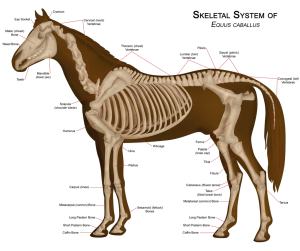
| Cannon bone | Horses | Equivalent to the human hand or foot bone extending from the wrist or ankle to the knuckles or toes
Bone extending from the carpus towards the fetlock This term is used on all four limbs |
| Fetlock | All | Equivalent to human joint between hand/foot bones and finger/toe bones
This term is used on all four limbs |
| Pastern | Horses | Length of the limb between the fetlock and the hoof, on all four limbs |
| Coffin bone | Horses | Bone equivalent to human’s most distal finger/toe bone
This bone is encapsulated within the hoof Term used in all four limbs |
| Flank | All | Lateral aspect of the caudal trunk of the body |
| Hook | Cows, sheep, goats | Cranial protruding aspect of the pelvis |
| Pin | Cows, sheep, goats | Caudal protruding aspect of the pelvis |
| Tail | All | Anatomy differs depending on species, but in general caudal extension of spine +/- extra hair |
| Dock | Sheep | Refers to a sheep’s tail that has been surgically removed |
| Stifle | All | Equivalent to human knee
Joint between the femur and the tibia/fibula (hind limb) |
| Hock/tarsus | All | Joint equivalent to human ankle
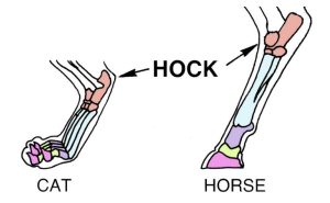
Tarsus is the name of the joint, but it is colloquially referred to as the “hock” in most animals (hind limb) |
| Dewclaw | Pigs, cows, sheep, goats | Two residual “toes” extending from the fetlock. Non-weight-bearing |
| Udder | Horses, cows, sheep, goats | Collection of mammary glands on the caudal-ventral portion of the body |
| Teat | All | Duct from the udder for milk to be excreted (nipple) |
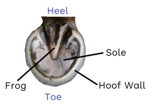
| Sole | All | Underside (palmar/plantar) aspect of the hoof
This term is used in all four limbs |
| Frog | Horse | Triangle of soft tissue on the underside of a horse’s hoof used for cushioning and balance |
Other species-specific terms you might hear include:
- Horns: Part of the integumentary system – hard protrusions from the heads of certain cows, goats, and sheep
- Wool: Specialized term for the thick curly hair of a sheep
Comparative Anatomy – Horses vs. Humans
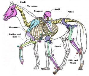
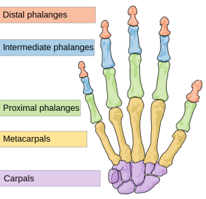
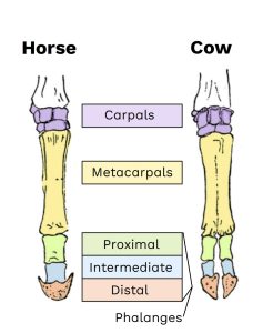
Animals and humans share the same general skeletal layout. In Figure 8.2, the bones are colour-coded to show how the skeletons compare.
Cattle
The following images will show the locations of the terms discussed above on both a diagram and a picture of a cow.
Organ locations
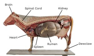
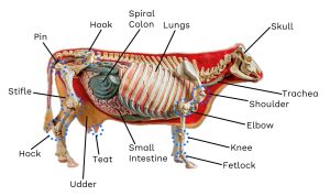
The internal organ systems were discussed in Chapters 3 and 4, so here is a model showing where those organs are located inside the body of a cow.
Sheep
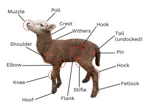
The following image will show the locations of the terms discussed above on an image of a lamb.
Goats
The following image will show the locations of the terms discussed above on an image of a goat.
Horses
The following image will show the locations of the terms discussed above on an image of a horse.
Skeletal Follow-Up
Pigs
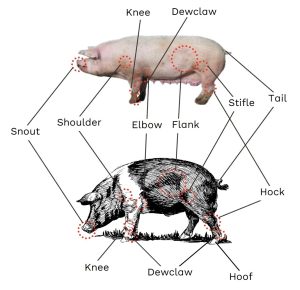
The following images will show the locations of the terms discussed above on both a diagram and a picture of a pig.
Exercises
Image Credits (images are listed in order of appearance)
Comparative view of skeletons of man and horse adapted by Mcapdevila, CC BY 3.0.
Scheme human hand bones by Mariana Ruiz Villarreal, CC0.
Various nail walkers’ toes by Ineuw, CC0.
Hock (PSF) by Pearson Scott Foresman, CC0.
Holsteiener BRW by Les Meloures, CC BY-SA 3.0.
Vintage Holstein cow by Unknown, CC0.
Modelo didatico bovino correto alt by Museum of Veterinary Anatomy FMVZ USP, CC BY-SA 4.0.
Didactic model of a cow by Museum of Veterinary Anatomy FMVZ USP, CC BY-SA 4.0.
Labelled sheep by Matéa David-Steel, NorQuest College. Used with permission.
Fluttering Bird, a Nigerian dwarf dairy goat by HeatherLion, CC BY-SA 3.0.
Tori horse universal by Rozpravka, CC0.
Sabot saint by Zorgglob, CC0.
Horseanatomy by WikipedianProlific, CC BY-SA 3.0.
Truie landrace by Zeilog, CC BY-SA 3.0.
Line art drawing of a hog by Unknown, CC0.
Overview of sheep and lamb in fence near Hostákov, Vladislav, Třebíč District by Frettiebot, CC0.
Haflinger horse on pasture in the Netherlands by Paula Jantunen, CC0.
An accurate description of the physical features of the anatomical parts of a species
Mammals with hooves

