6.5 Diagnostic Imaging Terminology
Overview
Diagnostic imaging is a large part of the diagnostic process of animal examinations. It is used to visualize and examine the internal structure of the animal’s body. Common imaging technology will be discussed in this chapter, but it is an ever-evolving field with new equipment and technology always in development.
Radiology
Radiology is the study of X-rays used for diagnostic purposes. Machines may be portable or stationary. Depending on their needs, clinics will vary in the type of equipment they use.
Radiographs or X-rays are a form of high energy radiation with a short wavelength, allowing it to penetrate through tissues. Because different tissues have different densities, they come up with different levels of opacity on an image, allowing us to visualize the interior of the animal.
Positioning of the patient is important in order to obtain a quality image, and knowledge of proper positioning techniques requires training. Once the animal is positioned, a series of images can be taken as needed. Projection is the path of the X-ray beam.
Review positional terms in directional and movement terms.
Example
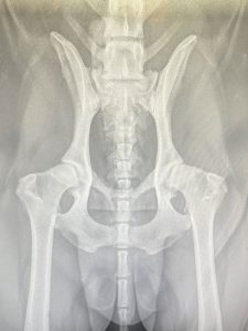
X-ray film is the picture of the image taken, traditionally reproduced on special material allowing the film to develop or process. Most clinics and facilities are setup for digital imaging, making it easier to diagnose, view, and send to other clinics.
Computed Tomography (CT) Scan
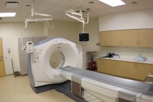
CT scan is an imaging technique that uses ionizing radiation to take several cross-sectional images. These images are put together by a computer which displays all the cross-sections together as a 3-D image. Contrast medium is often used to enhance the images.
CT scans are available at specialty veterinary facilities.
Magnetic Resonance Imaging (MRI)
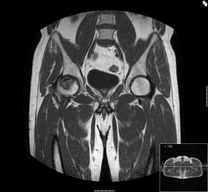
MRI is a medical imaging technique that passes radio waves and magnetic fields through the patient to create 3D images of the internal body structures. MRIs do not use X-rays, making them a safe procedure.
MRIs are available at specialty veterinary facilities.
Example
Ultrasonography or Ultrasound (U/S)
U/S is an imaging technique that uses the transmission of high-frequency sound waves into the body to generate an echo signal of high frequency sound waves, which a computer then converts into an image.
Ultrasound is common in many clinics. It is a safe option, including for pregnancy.
Endoscopy
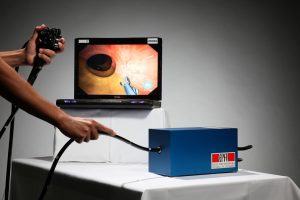
Endoscopy is the process of visually examining a hollow body organ or structure. Endoscopy falls into diagnostics when a DVM is exploring within the organs or structures for abnormalities, but it can also be considered surgical. Once they have explored the organs or structures, they can biopsy areas, which means they cut into organs for tissue samples. They may also remove foreign material, for example if a dog has eaten a toy, endoscopy tools could be used to remove it.
Imaging Safety
Safety of personnel is of utmost importance. Special gowns and gloves must be worn during X-rays.
Dosimetry is the measurement and determination of the amount of exposure to radiation.
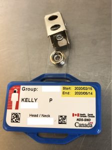
Dosimeter is a film badge worn by all staff when taking x-rays. It records the amount of exposure to the radiation generating equipment.
Diagnostic and Imaging Terms
Radiographic contrast medium: A substance used to enhance the image and structures that are typically difficult to see on a regular image
Barium sulfate: An example of a contrast medium. Other iodinated contrasts are available and widely used within the clinic
Exercise
Attribution
Unless otherwise indicated, material on this page has been adapted from the following resource:
OpenStaxCollege. (2013). Anatomy & physiology. Rice University. https://pressbooks-dev.oer.hawaii.edu/anatomyandphysiology/
Image Credits (images are listed in order of appearance)
Hip x-ray by Kelly Robertson, NorQuest College. Used with permission.
UPMCEast CTscan by daveynin, CC BY 2.0.
Femurkopfnekrose by Hermannthomas, CC0.
CSIRO ScienceImage 11130 CSIROs colonoscopy simulator developed using the latest computer gaming technology by division, CSIRO, CC BY 3.0.
Dosimeter by Kelly Robertson, NorQuest College. Used with permission.

