6.4 Laboratory Terminology
Overview
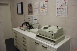
Laboratory testing is an important part of the patient evaluation process to determine health, establish a diagnosis, and monitor response to treatments and/or procedures. Specific terminology is used in the laboratory to describe tests and test results. Most practices have a basic lab within their regular clinic. We use the term “in-house” to describe tests done in the clinic. There are also laboratories outside of the clinical setting that we can send samples to for testing. Generally, these external labs can run more comprehensive tests than are available in-house. The term “off-site” laboratory is used to describe the labs outside of the regular clinic.
Routine Lab Tests
Many of the lab tests done in the clinic are examined under the microscope. Different samples will have different handling procedures, but all samples should be properly labelled, whether collected in-clinic or dropped off by a client.
Labelling requirements for samples:
- Owner’s name
- Patient’s name, DOB, species, and sex
- Date and time sample received (if collected and brought in by a client)
- Date and time sample collected
- Collection method (if relevant)
Blood Samples
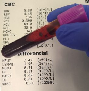
Blood is used in many of the laboratory tests done in the clinic. Blood is collected via venipuncture and proper animal restraint techniques are essential for obtaining a successful sample. Equipment required includes needles, syringes, collection tubes, alcohol, and bandages.
For more information about blood terms, see hematologic system.
Common Blood Tests
- Preanesthetic blood work: This test is run prior to anesthesia to ensure essential body organ systems like the liver and kidney are functioning properly.
- General/annual blood work: These tests are performed yearly with the wellness exam to track internal organ health.
- Geriatric blood work: Blood work to evaluate function of internal organs in senior animals; age 7–9+ years for most dogs and age 10–12+ years for most cats.
Blood Sample Abbreviations and Terms
White cell count: Number of leukocytes
Red cell count: Number of erythrocytes
Blood smear: Blood spread (and usually fixed and stained) on a microscope slide for examination under a microscope
PCV or hematocrit: Packed cell volume; percentage of red blood cells in the blood
CBC: Complete blood count; a collection of blood tests that count the different blood cells (RBCs, WBCs, platelets) and the different types of WBCs (neutrophils, lymphocytes, monocytes, eosinophils, basophils)
Chemistry panel: Groups of tests done using serum that measures liver and kidney enzymes along with other tests requested by the DVM, for example: glucose or thyroid levels
Urine Samples
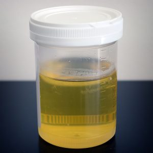
The routine urinalysis is a quick and relatively inexpensive test that can be readily performed in clinic or sent to off-site labs. Ideally, it should be completed with a yearly wellness exam. Often, a urinalysis occurs when there is a suspected urinary problem.
For more information about urine terms, see urinary system.
Urine Collection Methods
- Off the floor/out of litter box: Examination of urine from this route is not optimal, as it may contain bacteria or contaminants. Despite this knowledge, it may be the only method available to clients. This can serve as an initial test if no other route is possible.
- Free catch: This sample is obtained by catching the sample as it is released from the patient. When a dog is taken out for a walk, a clean collection container is held in the urine stream. Ideally, mid-stream urine is optimal. This type of sample involves the most stress-free collection method, but will still have the risk of bacteria and contamination during collection, which comes from the external genitalia.
- Urinary catheterization: This occurs by placing a sterile catheter directly into that bladder to obtain a sterile sample. It is mostly done on male dogs, as it is easier to perform compared to female dogs. Sedation on male dogs is not commonly needed, but female dogs and other species may need to be sedated to obtain a sample with this method.
- Cystocentesis: This method involves puncturing the bladder with a needle to obtain a sterile sample. This is the most sterile method to obtain a urine sample. It is relatively quick and easy to perform by the DVM or RVT, who are trained in this procedure.
atDove. (2016, December 16). Ultrasound guided cystocentesis [Video]. YouTube. https://youtu.be/-X_WsAlfTvc
Urinalysis
A urinalysis includes the following:
- Gross examination to assess colour, turbidity, and odour.
- Reagent strip test/dipstick test: A small test strip is dipped into the urine. These will change colour when positive.
- Common results analyzed are:
- Glucose: Sugar levels
- Ketones: Chemical in the body that results from the breakdown of fat
- pH: A measurement to determine acidity of the urine
- Blood: Presence of blood that might not be visualized under the microscope
- Common results analyzed are:
- Microscopic examination: Urine placed in a tube and centrifuged (spun at high speeds), then examined under a microscope
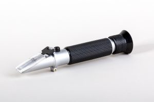
- A drop of urine is placed on a refractometer to read urine-specific gravity
Urine Sample Acronyms and Terms
Urine specific gravity (USG): Density or concentration of the urine. Performed with a refractometer.
- Clinical insight: USG is an important test used to determine kidney function. If the kidneys are not working, they cannot concentrate the urine!
Turbidity (of a liquid): Cloudy, opaque, or thick with suspended matter
GLU: Glucose
Fecal Testing
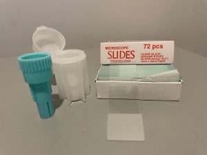
All species are at risk of internal parasites, intestinal bacterial, and viral infections. Some of these conditions can be identified by fecal examination. Samples can be obtained by collecting fecal material and testing it in the laboratory. Gross examination is inspecting the sample looking for consistency, colour, blood, mucus, and/or parasites. Microscopic examination is the next step of the fecal exam, with a direct smear. This involves placing a small amount of fecal material onto a slide for examination under the microscope. Given it is only a small area, sampled results may be inconclusive. The presence of endoparasites or parasite eggs (oocytes) can be tested with a few techniques. The mostly common is a fecal floatation, pictured in Fig 5.6. This method uses a sugar solution that allows the oocytes to float to the top of container. A glass cover slip is placed on top and taken to be examined under the microscope.
There are many more advanced fecal tests that can be performed at off-site laboratories to determine with better accuracy if internal parasites, bacteria, and viruses are present in the digestive tract.
Tissue and Fluid Samples
Tissue and fluid samples can be collected during surgery, local sedation, and necropsy. Samples can be of tissue or the fluid surrounding tissue, therefore collection tubes or containers may vary. Small samples may be placed in sterile culture tubes, while others may be placed in a sterile container alone or with formalin, depending on the type of sample and directions from the veterinarian. The cells of these samples will be examined.
Examples of tissue samples include skin and organs. Fluid samples include the substance from a mass, saliva, milk, joint fluid, and urine. Eye and ear material can also be sampled.
Biopsy: In laboratory terms, a biopsy means removing tissue for examination.
- An excisional biopsy is when the sample is removed in its entirety.
- An incisional biopsy is when the sample is cut into and a part is removed to be examined.
- A needle biopsy occurs when a needle is inserted into the tissue for examination.
- A punch biopsy uses a sterile circular blade that cuts into the tissue and pulls out a small sample.
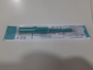
Skin scraping: Is obtained by using a scalpel and removing layers of skin for examination under the microscope
Cytology or Histology?
Cytology is the study of cells while histology is the study of tissue. Cytology is the test that is done on non-tissue samples (e.g. skin scraping, fluid, discharge), while histology is done on tissue biopsies.
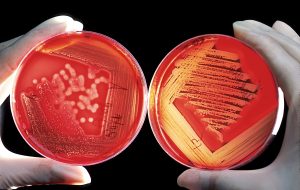
Cultures: A culture is a procedure done by taking a sample and allowing the microbes to reproduce in order to determine what type of bacteria or fungus they are. Most cultures are sent to an off-site lab for examination. The microbes take time to reproduce, so results are not immediate.
Culture and sensitivity: This test is similar to the culture test but includes a sensitivity component as well. Sensitivity testing determines which medications the sample is sensitive to, and therefore which will kill the microbes.
SNAP Tests
SNAP tests are rapid-result tests that use a small amount of blood or fecal matter (depending on the test) to determine presence of a certain disease. The tests in clinic will vary depending on geographical areas. Common SNAP tests are heartworm, parvo, FIV/FeLV combo, leptospirosis, and giardia.
Laboratory Abbreviations and Terms
PPE: Personal protective equipment
DOB: Date of birth
WNL: Within normal limits
TNTC: Too numerous to count
- Generally written after observing a slide under a microscope and a certain type of bacteria or cell is present in such high numbers that they are not counted.
Centrifuge: The centrifuge is a commonly used tool in our laboratory. It uses centrifugal force to separate substances in liquid or solid media according to particle size and density differences.
Direct smear: A sample of blood or feces placed directly onto a microscope slide for evaluation
Zoonosis: An infectious disease that can spread between animals and humans
Lumpectomy: Removal of a lump or mass
Excision: To cut or surgically remove
Laboratory Sample Overview: Common In-House Tests
| Lab Test | Samples | Results | Reasoning |
|---|---|---|---|
| Urinalysis (UA) | Urine | USG
Cells, crystals, bacteria pH, glucose |
Can help determine presence of UTI
Can help determine kidney function |
| Fecal Float and Direct Smears | Feces | Presence of parasites and bacteria | Determine presence of endoparasites |
| Blood work | Blood | PCV and TP
CBC and chemistry Morphology |
Determine function of internal organs, presence of disease, presence of bleeding, etc. |
| Cytology | Ear swabs
Vulva swabs FNA Skin scraping Impression smear Etc. |
Visualizes cellular population and presence of bacteria, and potentially ectoparasites and yeast
Results dependent on sample |
Helps diagnose ear infections, skin infections, masses, etc.
Reasoning dependent on sample |
Note: There are many other tests that are more specific. These are generally sent to an external laboratory.
Handling and Storage
Blood: Depending on the test, different tubes are required. Samples are run promptly or after clotting. If they cannot be tested immediately, they will be stored in the refrigerator.
Urine: Once the urine is collected, it should be analyzed within 30 minutes, otherwise it should be stored in the refrigerator and tested within 12 hours.
Feces: Giving clients a clean sealed container is optimal. Otherwise, once the sample is obtained, transferring it to a sealed container is ideal. Testing should occur immediately if possible or refrigerated and tested as soon as possible, to reduce the risk of bacterial growth.
Tissue and fluid samples: Most samples will be sent to off-site laboratories. Instructions provided by the lab where the sample is being sent for the specific test required should be followed for handling and storage.
Laboratory Safety
Safety for yourself and others is of the utmost importance in the clinic. Every clinic must follow and obey safety guidelines set out for veterinary clinics. Each clinic should have operating procedures for handling samples.
- Have all PPE ready prior to procedures.
- Know where and how to use eyewash station.
- Ensure all biohazard waste, such as urine, blood, and bodily fluids are in the appropriate labelled containers.
- All sharps are placed in a sharps container after use.
- Properly disinfect surfaces and instruments after each animal.
Exercise
Attribution
Unless otherwise indicated, material on this page has been adapted from the following resource:
Burton, E., & Lalande, A. (2021). Clinical veterinary diagnostic laboratory. University of Minnesota. https://pressbooks.umn.edu/cvdl/.
References
atDove (2016, December 16). Ultrasound guided cystocentesis [Video]. Youtube. https://youtu.be/-X_WsAlfTvc
Image Credits (images are listed in order of appearance)
Veterinary lab by Sujalajus, CC BY-SA 3.0.
Complete blood count and differential by SpicyMilkBoy, CC BY-SA 4.0.
Urine sample by Turbotorque, CC0.
Portable refractometer by CEphoto, Uwe Aranas, CC BY-SA 3.0.
Fecal test by Kelly Robertson, NorQuest College. Used with permission.
Disposable biopsy punch by Ajay Kumar Chaurasiya, CC BY-SA 4.0.
Agarplate redbloodcells by Bill Branson, CC0.
withdrawing blood from a vein.
the liquid portion of the blood after it has clotted.
is a panel of medical tests that includes physical (macroscopic) examination of the urine
cloudiness
an instrument that measures the density of urine compared to pure water via the refractive index.
Parasites living within the tissues and organs of the host.
post-mortem (after death) examination on an animal species.
also called infectious waste or biomedical waste, is any waste containing infectious materials or potentially infectious substances such as blood.

