
1 Red Blood Cell Maturation
Michelle To and Valentin Villatoro
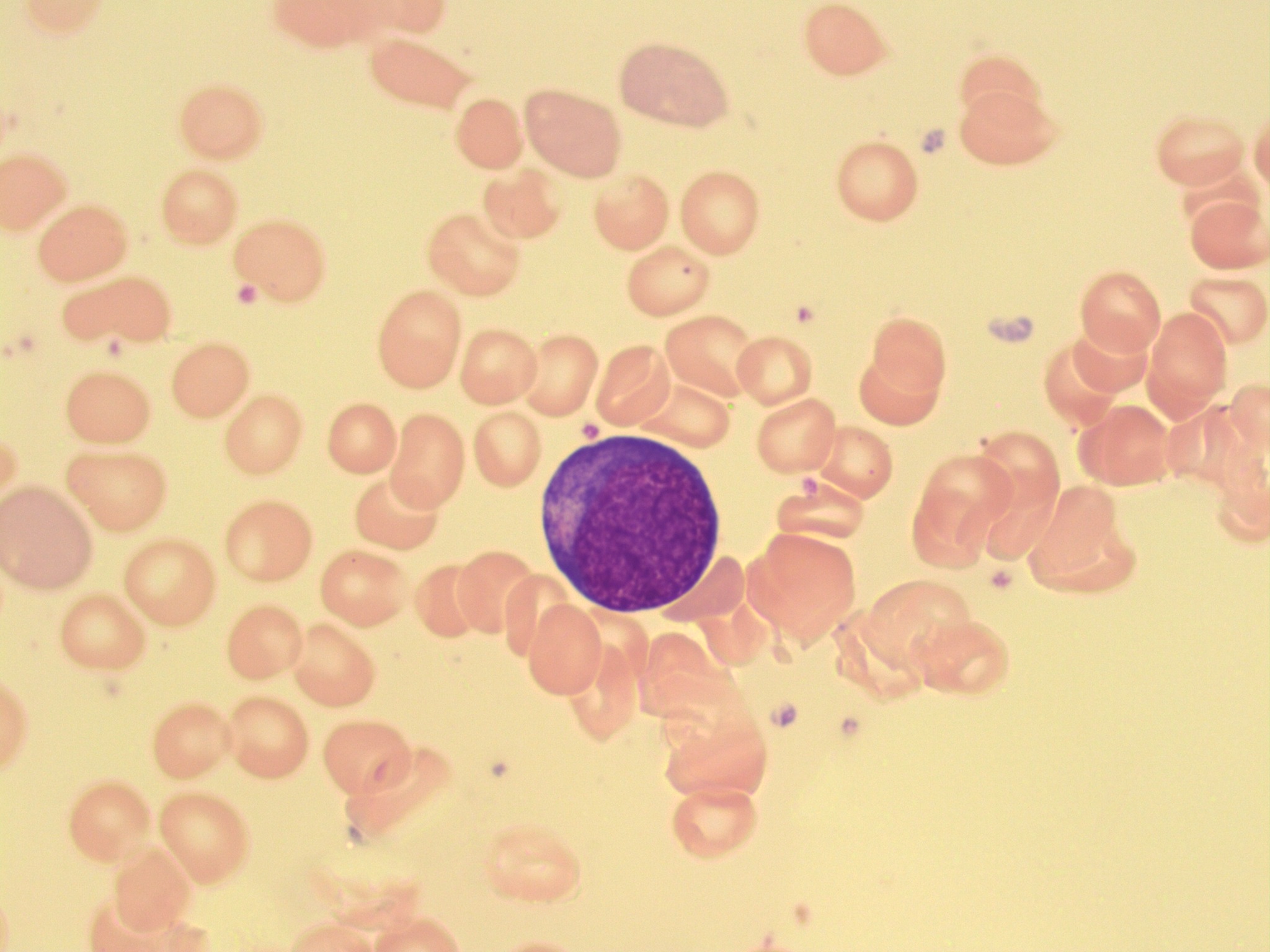
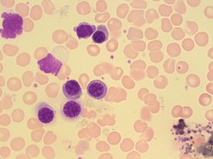
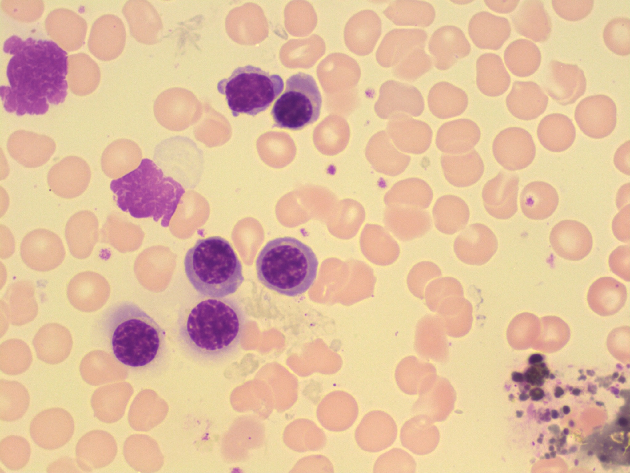
Pronormoblast (Rubriblast, Proerythroblast)
-

- Image shows a pronormoblast in a bone marrow smear. 100x oil immersion. From MLS Collection, University of Alberta, https://doi.org/10.7939/R3S46HN49
-
- Image taken from a bone marrow smear shows a pronoroblast in the center. 100x oil immersion. From MLS Collection, University of Alberta, https://doi.org/10.7939/R3KW5803R

Notes: Largest of the RBC maturation series 1
Nucleus-to-Cytoplasm Ratio: 8:1 (High) 1,2
Nucleoli: 0-2 2,3
Nucleus:1-3
Round to oval, central
Fine, homogeneous chromatin
Reddish-blue colour under Wright stain
Cytoplasm:1-3
Small to moderate amount of cytoplasm
Dark blue cytoplasm (due to large RNA content)
Golgi may be seen (pale area next to the nucleus)
% in Bone Marrow: 1% 1-3
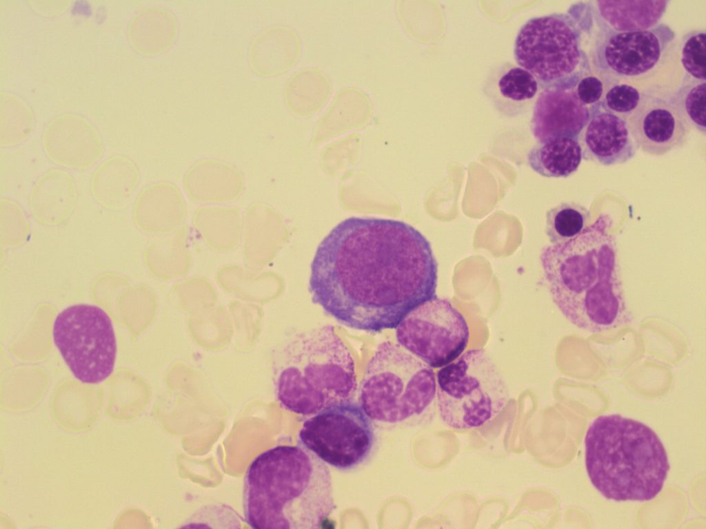
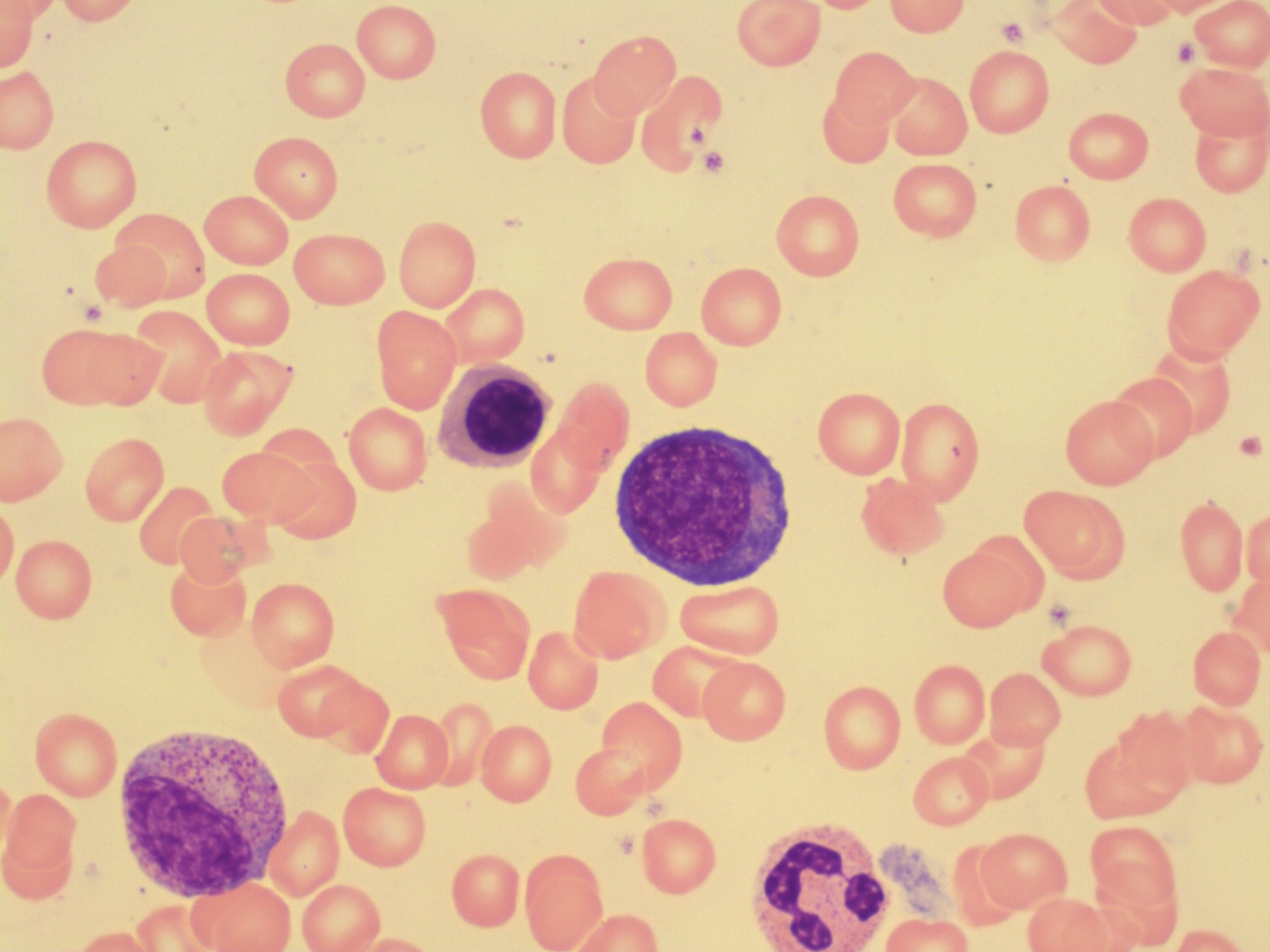
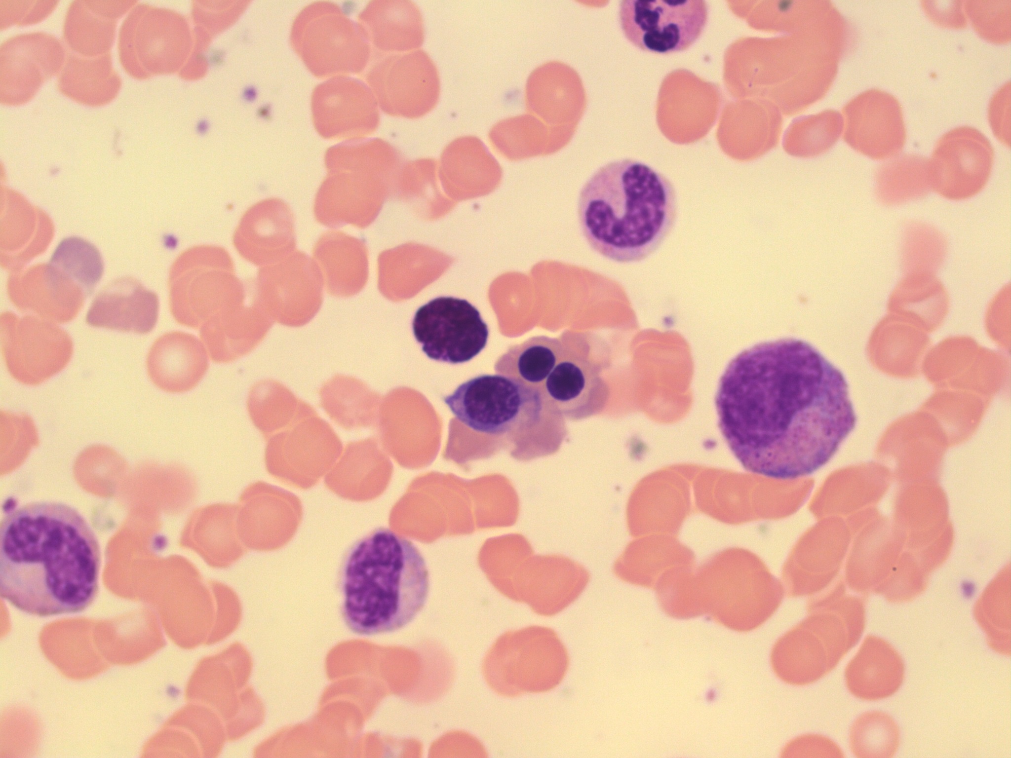
Basophilic Normoblast (Prorubricyte, Basophilic Erythroblast)
-

- Image taken from a bone marrow smear shows a basophilic normoblast (center top). A metameylocyte is present on the right of the cell. From MLS Collection, University of Alberta, https://doi.org/10.7939/R3PC2TQ99
-
- Image taken from a bone marrow smear shows a basophilic normoblast in the center. 100x oil immersion. From MLS Collection, University of Alberta, https://doi.org/10.7939/R38C9RK46
-
- Image taken from a bone marrow smear shows a basophilic normoblast in the center. 100x oil immersion. From MLS Collection, University of Alberta, https://doi.org/10.7939/R3HX1662B
Notes: Smaller than Pronormoblasts 3
Nucleus-to-Cytoplasm Ratio: 6:1 1
Nucleoli: 0-1 2
Nucleus: 1-3
Round to slightly oval, central
Chromatin is coarser and slightly clumped
Dark violet in colour
Indistinct nuclei or not visible
Cytoplasm: 1,2
Dark blue (due to large RNA content)
May see a perinuclear halo (unstained mitocondria)
May have a slight pink tinge due to the production of hemoglobin
% in Bone Marrow: 1-5% 3
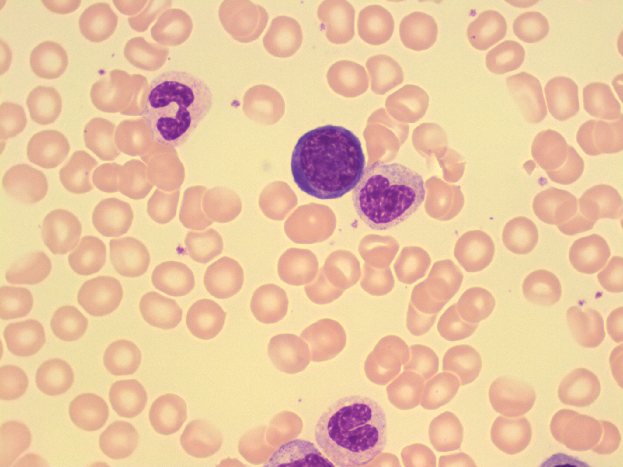
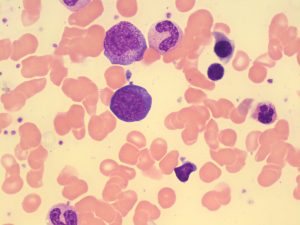
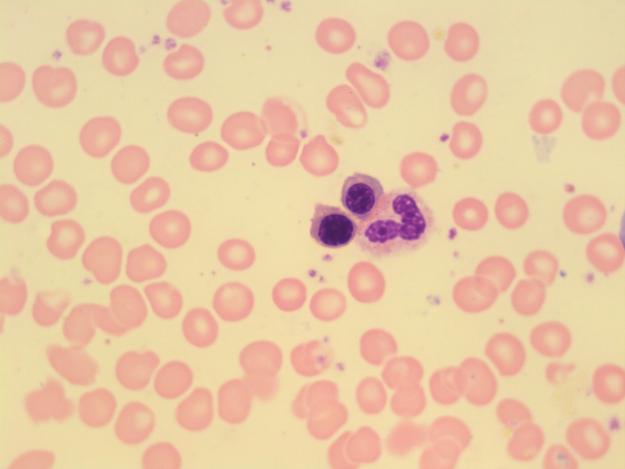
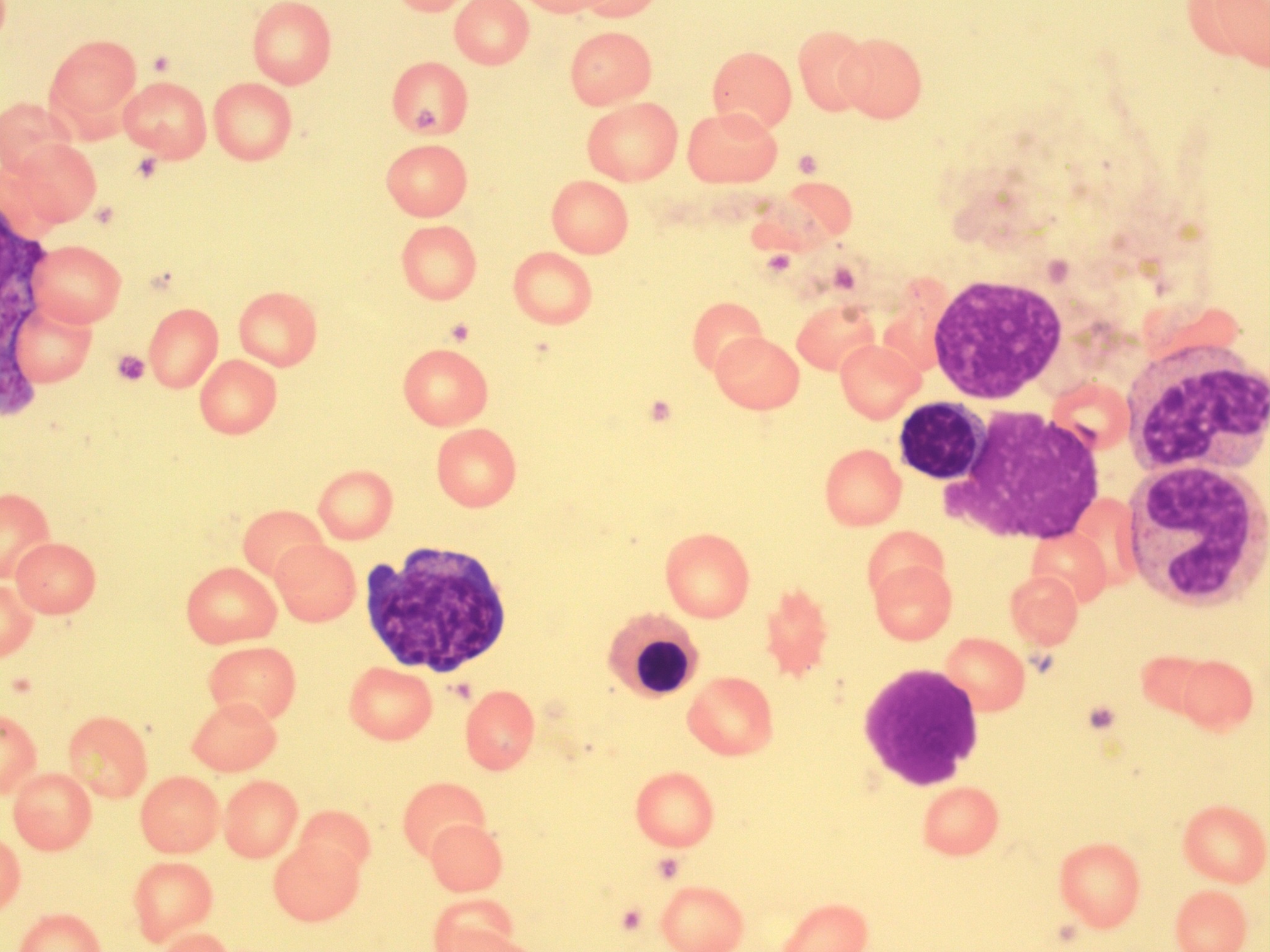
Polychromatic Normoblast (Rubricyte, Polychromatic Erythroblast)
-
- Image taken from a bone marrow smear demonstrating two polychromatic normoblasts in the center. 100x oil immersion. From MLS Collection, University of Alberta, https://doi.org/10.7939/R3C53FG8N
-
- An image taken from a bone marrow smear showing two polychromatic normoblasts (left) beside a neutrophil (right). 100x oil immersion. From MLS Collection, University of Alberta, https://doi.org/10.7939/R3MP4W36V
-
- Image taken from a bone marrow smear showing multiple polychromatic normoblasts. 100x oil immersion. From MLS Collection, University of Alberta, https://doi.org/10.7939/R31J97Q0D
Notes: Last RBC maturation stage capable of mitosis 1
Nucleus-to-Cytoplasm Ratio: 4:1 1-3
Nucleoli: None 2
Nucleus: 1-3
Round, eccentric
Chromatin is coarse, irregularly clumped
Cytoplasm: 1,2
Abundant
Gray-blue to pink (due to hemoglobin production and RNA content)
% in Bone Marrow: 5-30% 3
% in Peripheral Blood: Normally NOT present in the peripheral blood but some may be seen in the peripheral blood smears of newborns.3
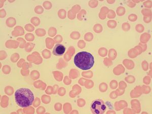
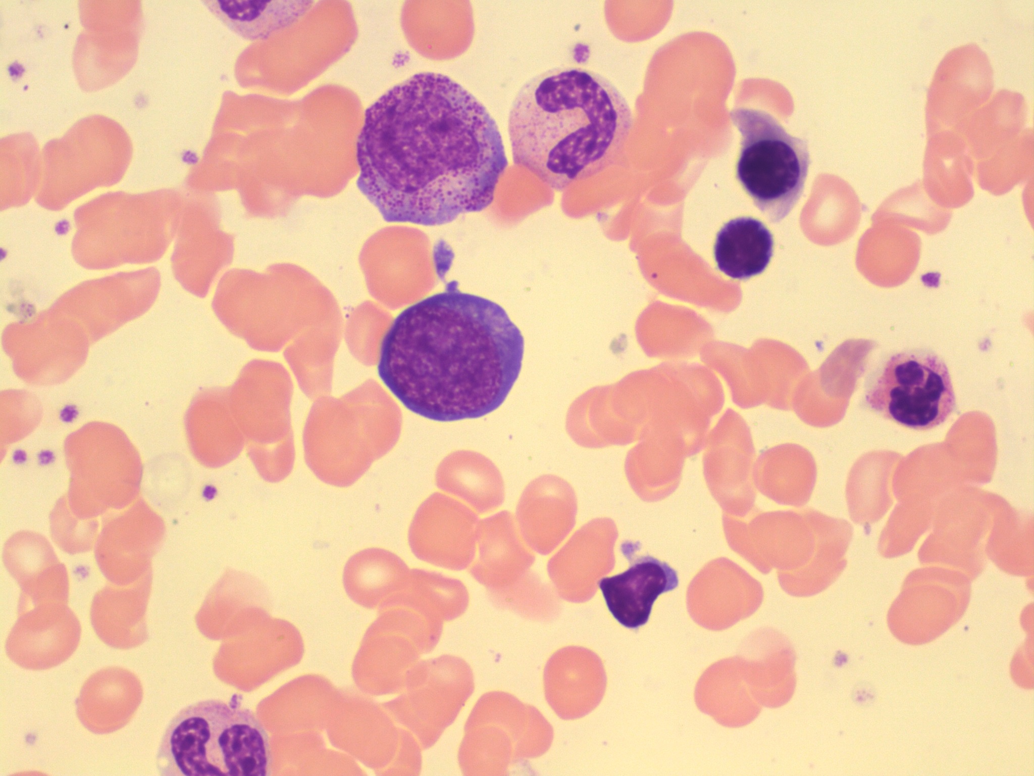
Orthochromic Normoblast (Metarubricyte, Orthochromatic Erythroblast)
-

- Image taken from a bone marrow smear showing two orthochromic normoblasts in the center right. Note the dark staining nucleus and condensed chromatin. 100x oil immersion. From MLS Collection, University of Alberta, https://doi.org/10.7939/R30Z71C0H
-
- Image taken from a bone marrow smear showing an orthochromic normoblast in the center bottom. Note the dark staining nucleus and condensed chromatin. 100x oil immersion. From MLS Collection, University of Alberta, https://doi.org/10.7939/R3W669Q5T
-
- An image from a bone marrow smear showing an orthochromic normoblast (center) ejecting its condensed nucleus. 50x oil immersion. From MLS Collection, University of Alberta, https://doi.org/10.7939/R3599ZH3P

Notes: The smallest RBC precursor and incapable of further DNA synthesis at this stage.3
Nucleus-to-Cytoplasm Ratio: 1:1 (Low) 3
Nucleoli: None 2-3
Nucleus: 1,2
Round, eccentric
Fully condensed chromatin with pyknotic features
Cytoplasm: 1,2
Pink or salmon; May appear slightly blue due to residual RNA
% in Bone Marrow: 5-10% 2
% in Peripheral Blood: Normally NOT present in the peripheral blood but some may be seen in the peripheral blood smears of newborns. 3
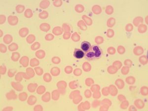
Reticulocyte (Polychromatic Erythrocyte, Diffusely Basophilic Erythrocyte)
-
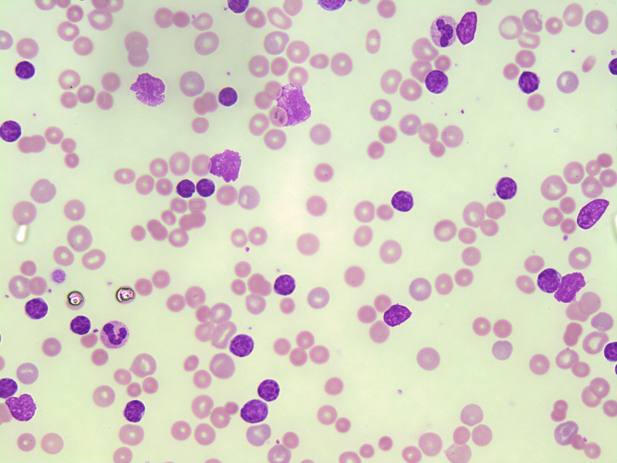
- A peripheral blood smear image representing hereditary spherocytosis. Marked polychromasia is present representing increased amounts of reticulocytes. 50x oil immersion. From MLS Collection, University of Alberta, https://doi.org/10.7939/R3N873F1Q
-
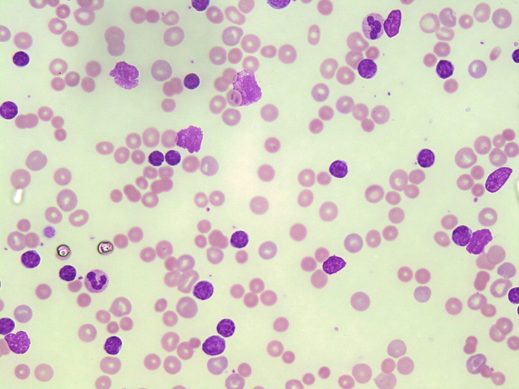
- A image of a CLL peripheral blood smear showing polychromasia in numerous red blood cells. The polychromasia represents reticulocytes. 50x oil immersion. From MLS Collection, University of Alberta, https://doi.org/10.7939/R3513VB2P
-

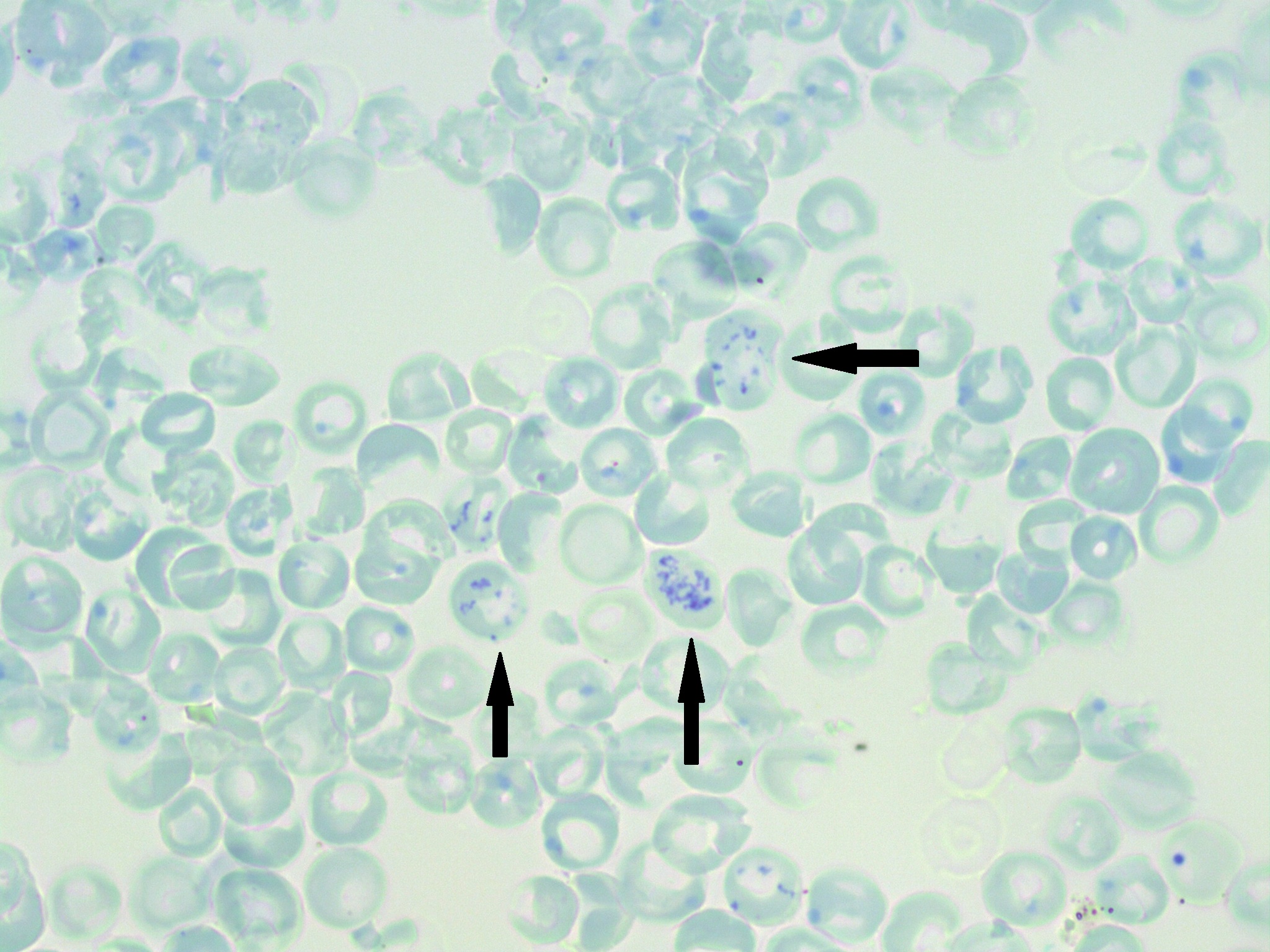
- A supravital stained peripheral blood smear showing multiple reticulocytes (indicated by arrows). Note the blue stained reticulum resembling “beads on a string”. 100x oil immersion. From MLS Collection, University of Alberta, https://doi.org/10.7939/R31G0J94K
-

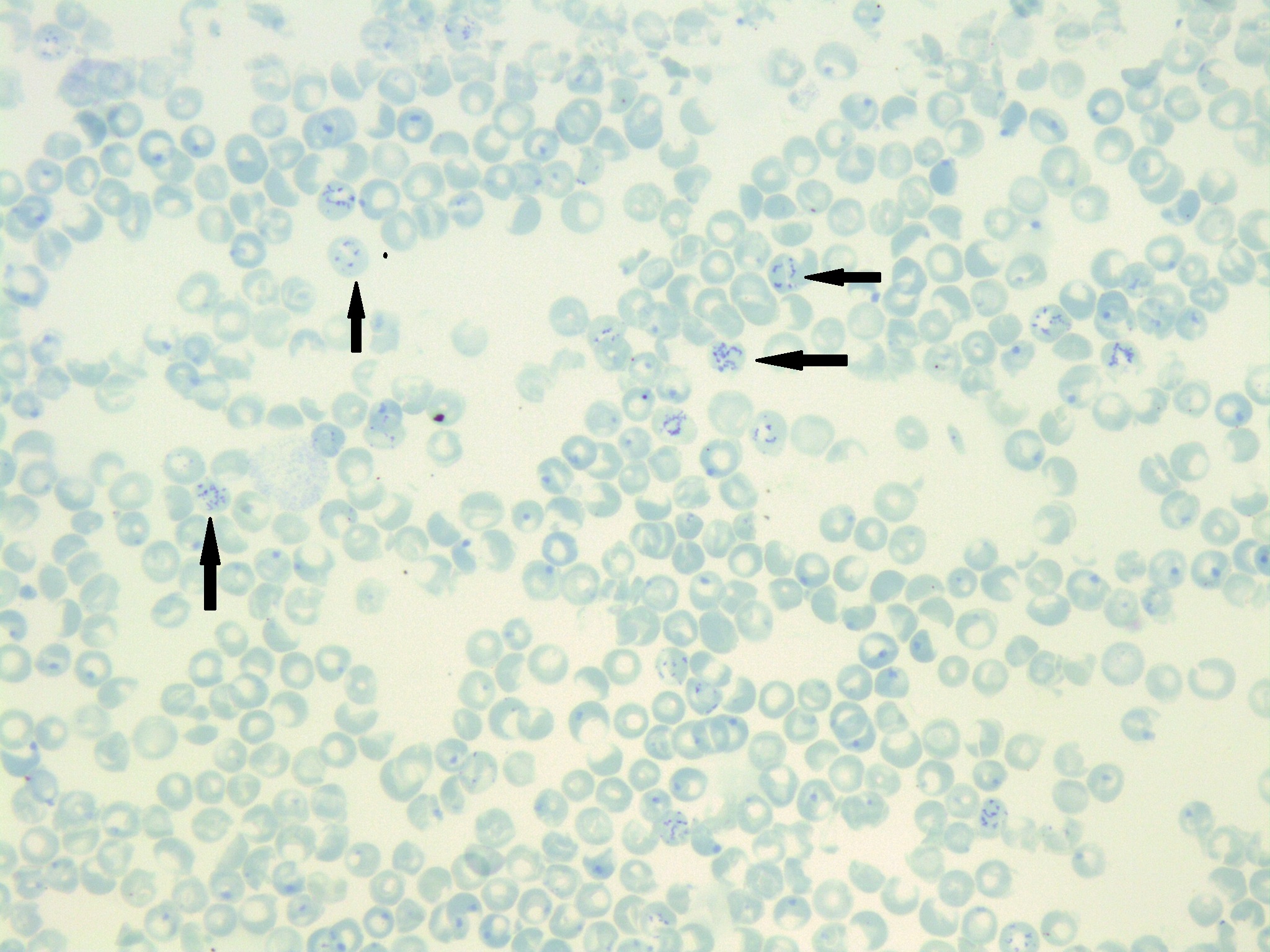
- An image from a peripheral blood smear stained with a supravital stain showing Heinz Body inclusions (large single blue inclusions) and reticulocytes containing dark-blue linear chains of granulation. New methylene blue. 50x oil immersion. From MLS Collection, University of Alberta, https://doi.org/10.7939/R3WP9TN91
Notes: the nucleus has now been expelled from the cell, residual RNA gives the cell a polychromatic appearance. The use of supravital stains can help to identify and enumerate Reticulocytes by visualizing reticular inclusions (linear granulation, with a “beads on a string” appearance, see figure below). (Har ch 1 pg 13)
Nucleus-to-Cytoplasm Ratio: N/A 2
Nucleoli: N/A 2
Nucleus: N/A 2
Cytoplasm: 2,3
Light blue-purple to pink (due to residual RNA content and high hemoglobin content)
% in Bone Marrow: 1% 2
% in Peripheral Blood: 0.5-2% 2
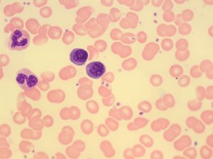
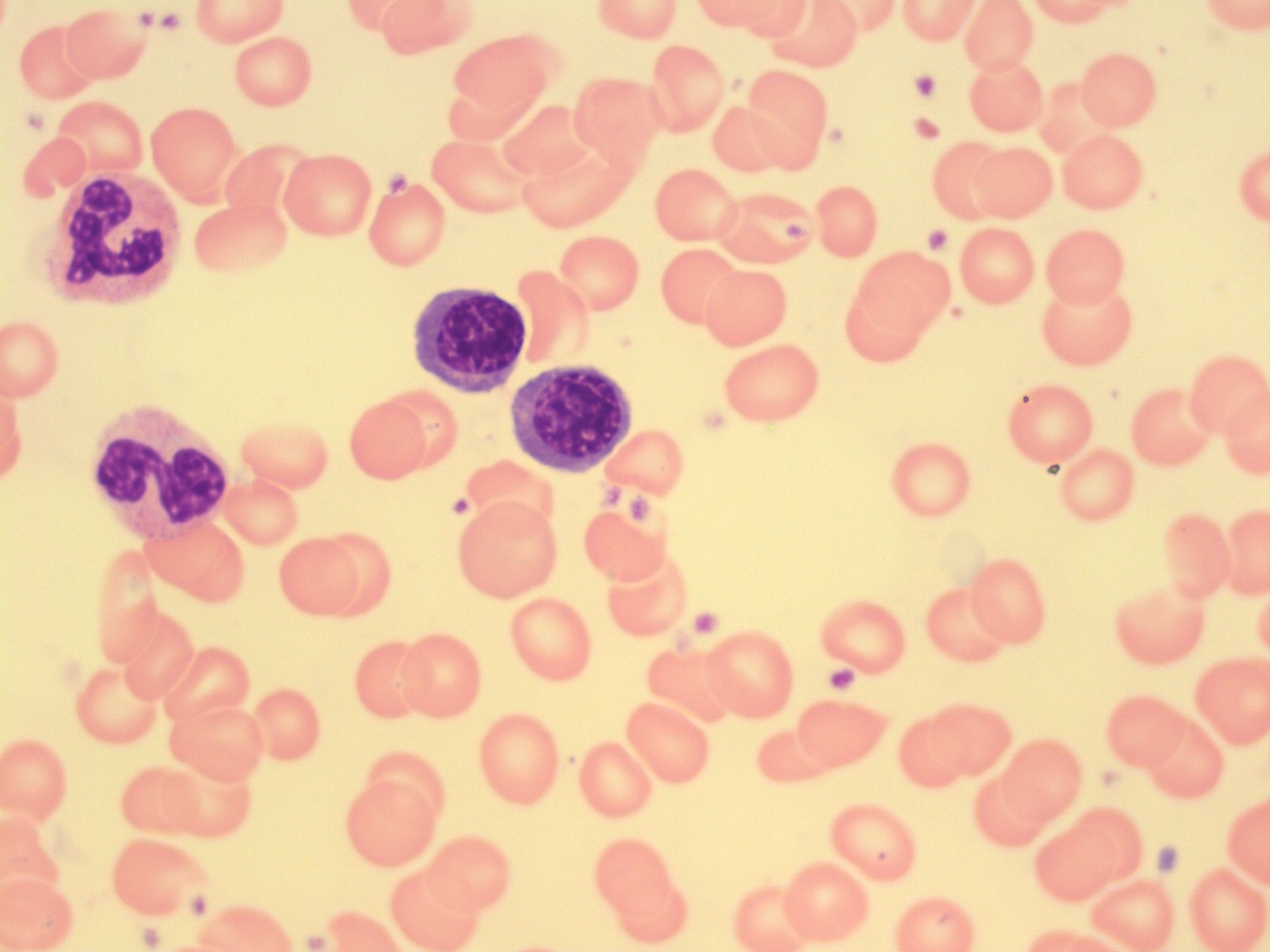
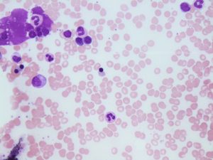
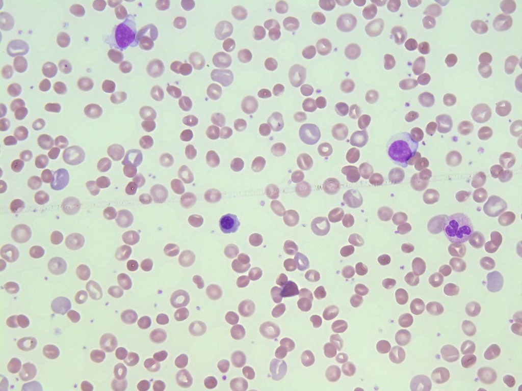
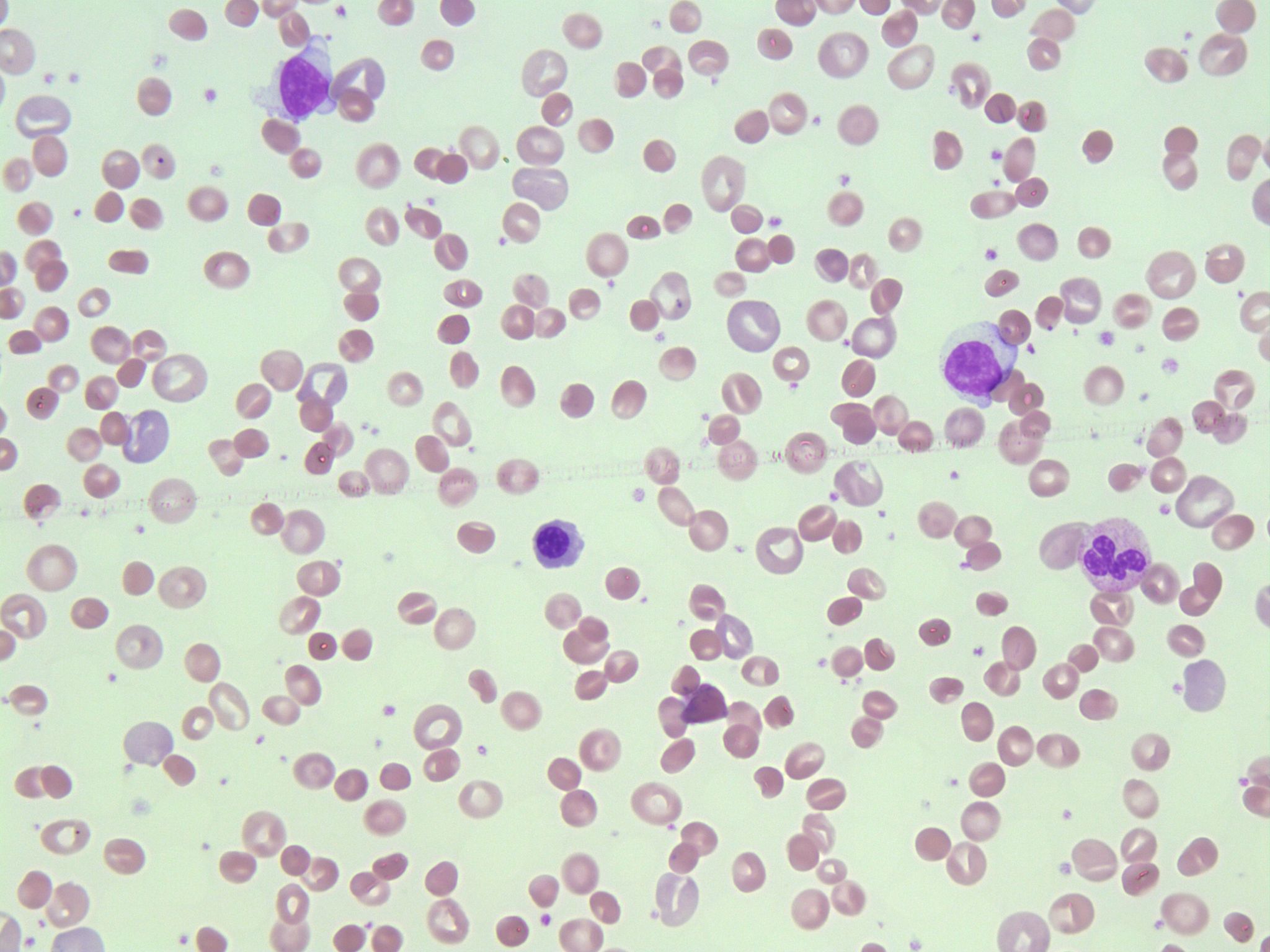
Erythrocyte (Discocyte)
-
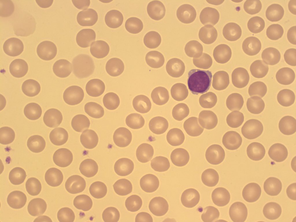
- An image from a peripheral blood smear showing normochromic, normocytic red blood cells. A small lymphocyte is present from comparison. 100x oil immersion. From MLS Collection, University of Alberta, https://doi.org/10.7939/R3W669Q69

-
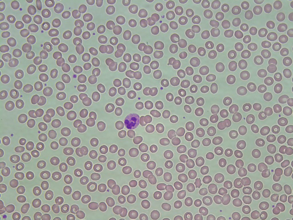
- An image from a peripheral blood smear showing normal mature erythrocytes. A neutrophil is present for size comparison. 50x oil immersion. From MLS Collection, University of Alberta, https://doi.org/10.7939/R32J68K44
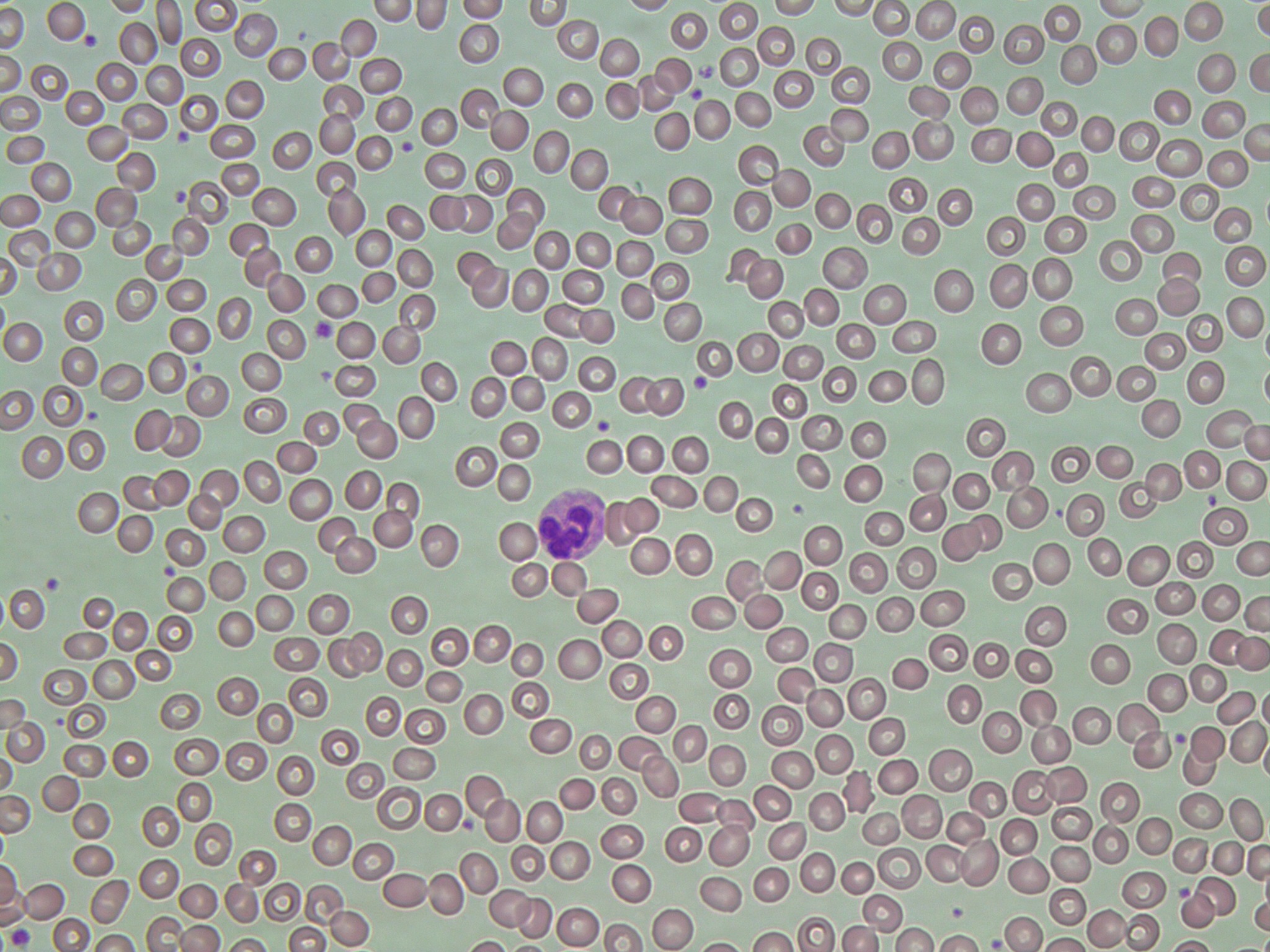
Notes: The mature red blood cell is biconcave in shape and lacks ribosomes and mitochondria; therefore, it lacks the ability to synthesize proteins such as hemoglobin and enzymes such as G6PD.1
Nucleus-to-Cytoplasm Ratio: N/A 2
Nucleoli: N/A 2
Nucleus: N/A 2
Cytoplasm: 2-3
Pink-salmon colour with an area of central spanning one-third of the diameter. Cell should contain no inclusions.
% in Bone Marrow: N/A 2
% in Peripheral Blood: Predominant 2
References:
1. Robinson S, Hubbard J. The erythrocyte. In: Clinical laboratory hematology. 3rd ed. New Jersey: Pearson; 2015. p. 59-76.
2. Rodak BF, Carr JH. Erythrocyte maturation. In: Clinical hematology atlas. 5th ed. St. Louis, Missouri: Elsevier Inc.; 2017. p. 17-30
3. Bell A, Harmening DM, Hughes VC. Morphology of human blood and marrow cells. In: Clinical hematology and fundamentals of hemostasis. 5th ed. Philadelphia: F.A. Davis Company; 2009. p. 1-41.
