
60 Megakaryocytes
Michelle To and Valentin Villatoro
-

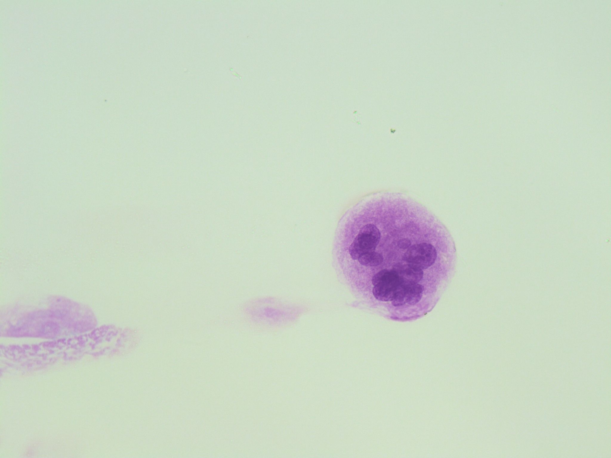
- An image from a bone marrow smear showing a normal megakaryocyte with multiple nuclear lobes. 50x oil immersion. From MLS Collection, University of Alberta, https://doi.org/10.7939/R3FF3MF8N
-
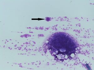
- An image from a bone marrow smear showing a megakaryocyte (indicated by an arrow) in the tails of the smear. 10x magnification. From MLS Collection, University of Alberta, https://doi.org/10.7939/R3TQ5RV93

-
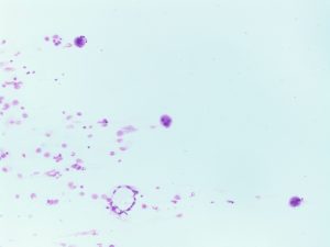
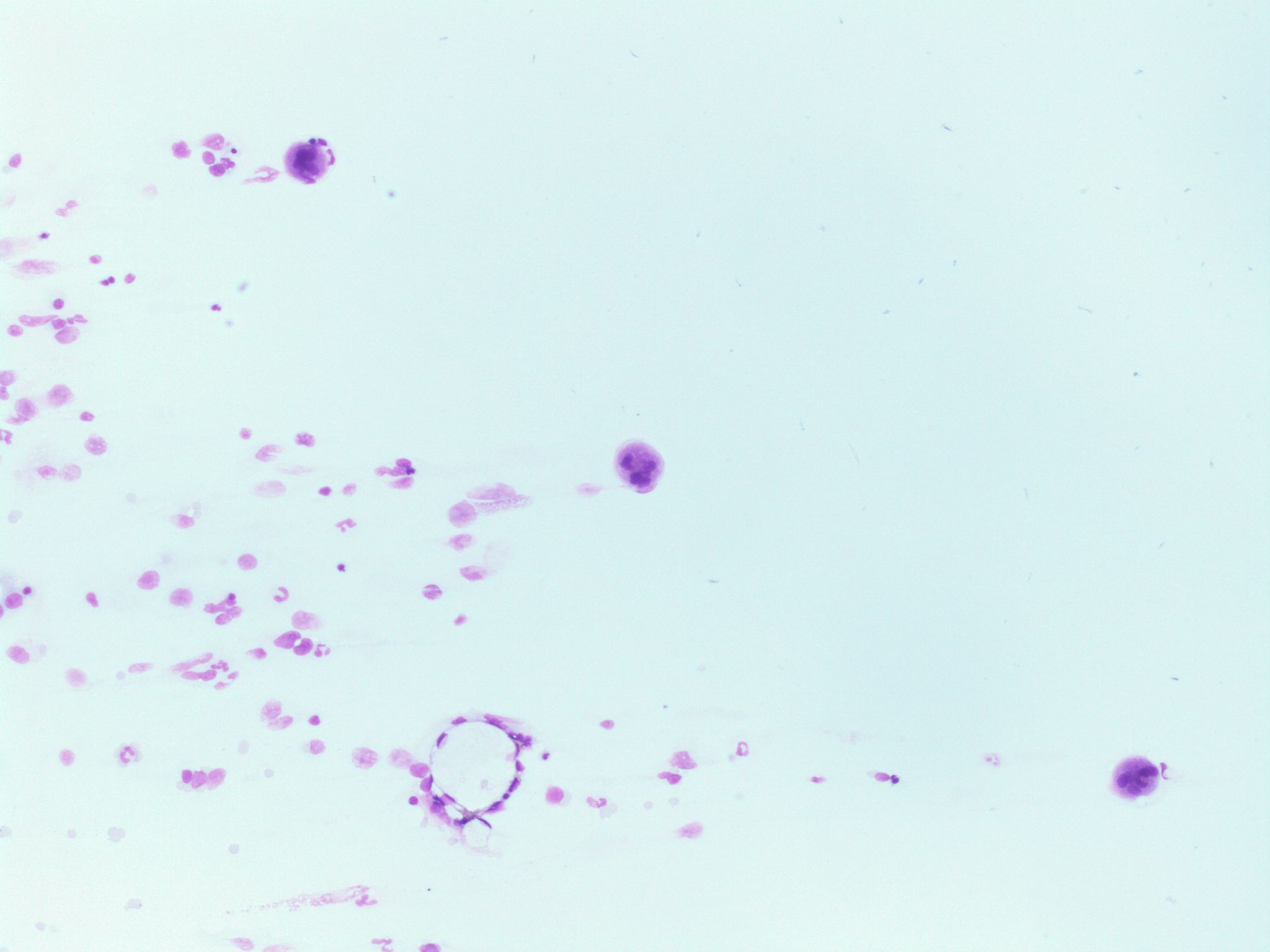
- An image from a bone marrow smear showing three megakaryocytes in the tail of the smear. 10x magnification. From MLS Collection, University of Alberta, https://doi.org/10.7939/R3K64B82D
Notes: Develop and are mainly found in the bone marrow. Maturation usually involves the division of nucleus but not the division of the cytoplasm, this gives rise to a polyploid cell.1
Nucleus-to-Cytoplasm Ratio: Variable 2
Nucleoli: N/A 2
Nucleus:
Variable number of lobes (2-32)2
Cytoplasm:2
Abundant
Blue to purple cytoplasm
Reddish blue granules may be visible
% in Bone Marrow: 5-10 (per field at 100x magnification)2
% in Peripheral Blood: None
References:
1. Lynne Williams J. The Platelet. In: Clinical laboratory hematology. 3rd ed. New Jersey: Pearson; 2015. p. 144–53.
2. Rodak BF, Carr JH. Megakaryocyte maturation. In: Clinical hematology atlas. 5th ed. St. Louis, Missouri: Elsevier Inc.; 2017. p. 31-40.
