
59 Macrophages
Michelle To and Valentin Villatoro
-
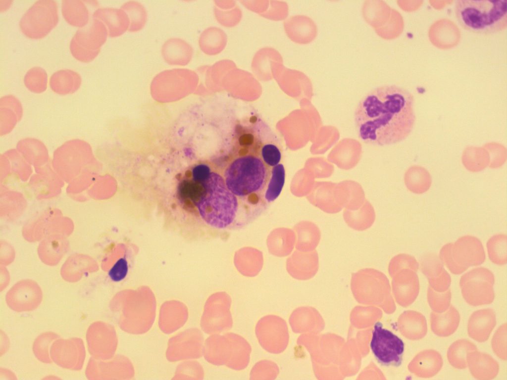
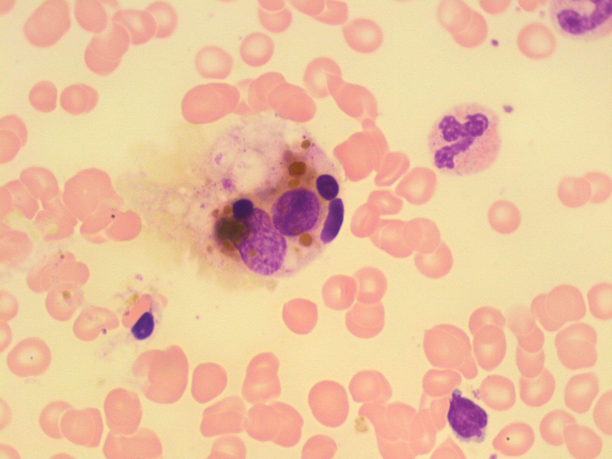
- An image from a bone marrow smear showing a macrophage with a valuolated and granular cytoplasm. 100x oil immersion. From MLS Collection, University of Alberta, https://doi.org/10.7939/R3DV1D40B
-
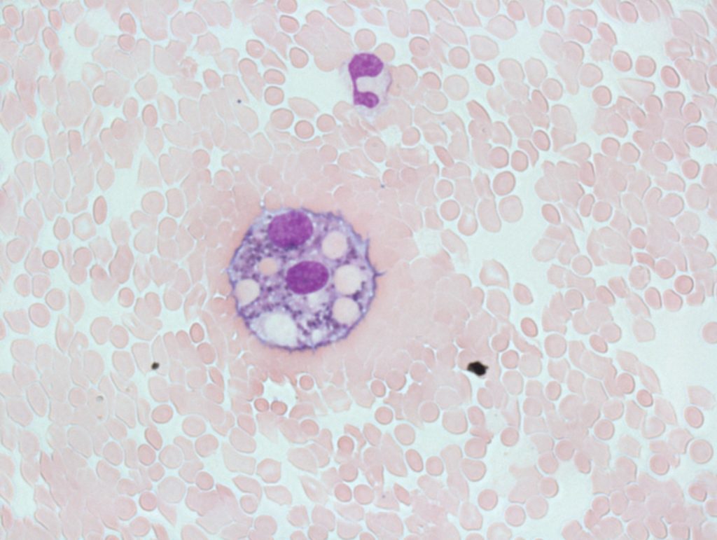
- An image from a Cerebrospinal Fluid (CSF) cytospin slide showing erythrophagocytosis in a macrophage. Ingested red blood cells, vacuolation, and hemosiderin granules can be seen within the cell. 60x oil immersion. From MLS Collection, University of Alberta, https://doi.org/10.7939/R36H4D570

Notes: Macrophages represent the mature form of monocytes when they leave the circulation and enter the tissues.1
Nucleus-to-Cytoplasm Ratio: N/A 2
Nucleoli: 1-2 2
Nucleus:2
Variable shapes (kidney, rounded, indented, oval)
Eccentrically located
Dark purple, coarse, clumped chromatin
Cytoplasm:1,2
Abundant
Irregular shaped
Many azurophilic granules
May contain ingested material and/or storage granules (hemosiderin, red blood cells, lipids, microorganisms, debris)
May contain vacuoles
References:
1. Landis-Piwowar K. Granulocytes and Monocytes. In: Clinical laboratory hematology. 3rd ed. New Jersey: Pearson; 2015. p. 97-121.
2. Rodak BF, Carr JH. Monocyte maturation. In: Clinical hematology atlas. 5th ed. St. Louis, Missouri: Elsevier Inc.; 2017. p. 55-64.
