
55 Granulocytes and Granulocyte Maturation
Michelle To and Valentin Villatoro
Myeloblast/Blast
-
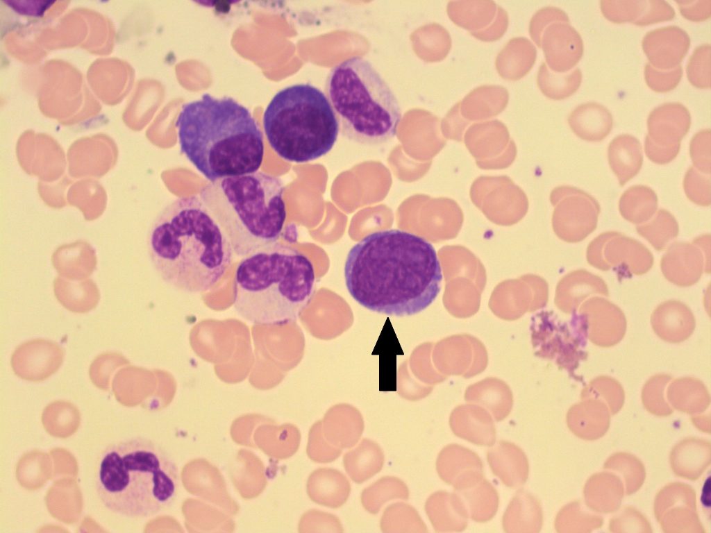
- An image from a bone marrow smear with a blast (indicated with an arrow). 100x oil immersion. From MLS Collection, University of Alberta, https://doi.org/10.7939/R3JM23X5J
-
- An image from a bone marrow smear with a blast (indicated with an arrow). 50x oil immersion. From MLS Collection, University of Alberta, https://doi.org/10.7939/R3JM23X5J
Notes: Earliest distinguishable and recognizable stage of granulocyte maturation.1
Nucleus-to-Cytoplasm Ratio: 4:1 2
Nucleoli: 1-51
Nucleus:1,3
Round to oval
Central or eccentrically located
Loose, open, evenly stained, reddish-purple, chromatin
Cytoplasm:1,2
Dark to light basophilia
May contain granules (up to 20)
Golgi may be seen (pale area next to the nucleus)
Normal % in Bone Marrow: 0-2%2
Normal % in Peripheral Blood: 0%2
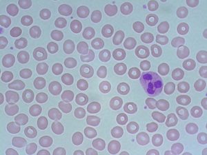
Promyelocyte
-
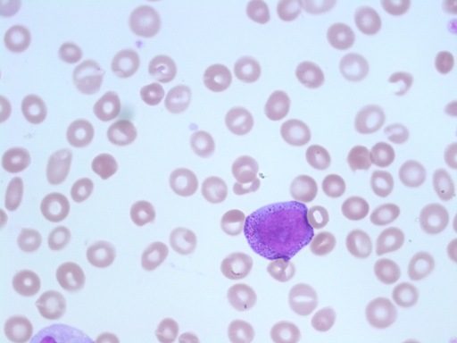
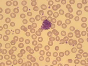
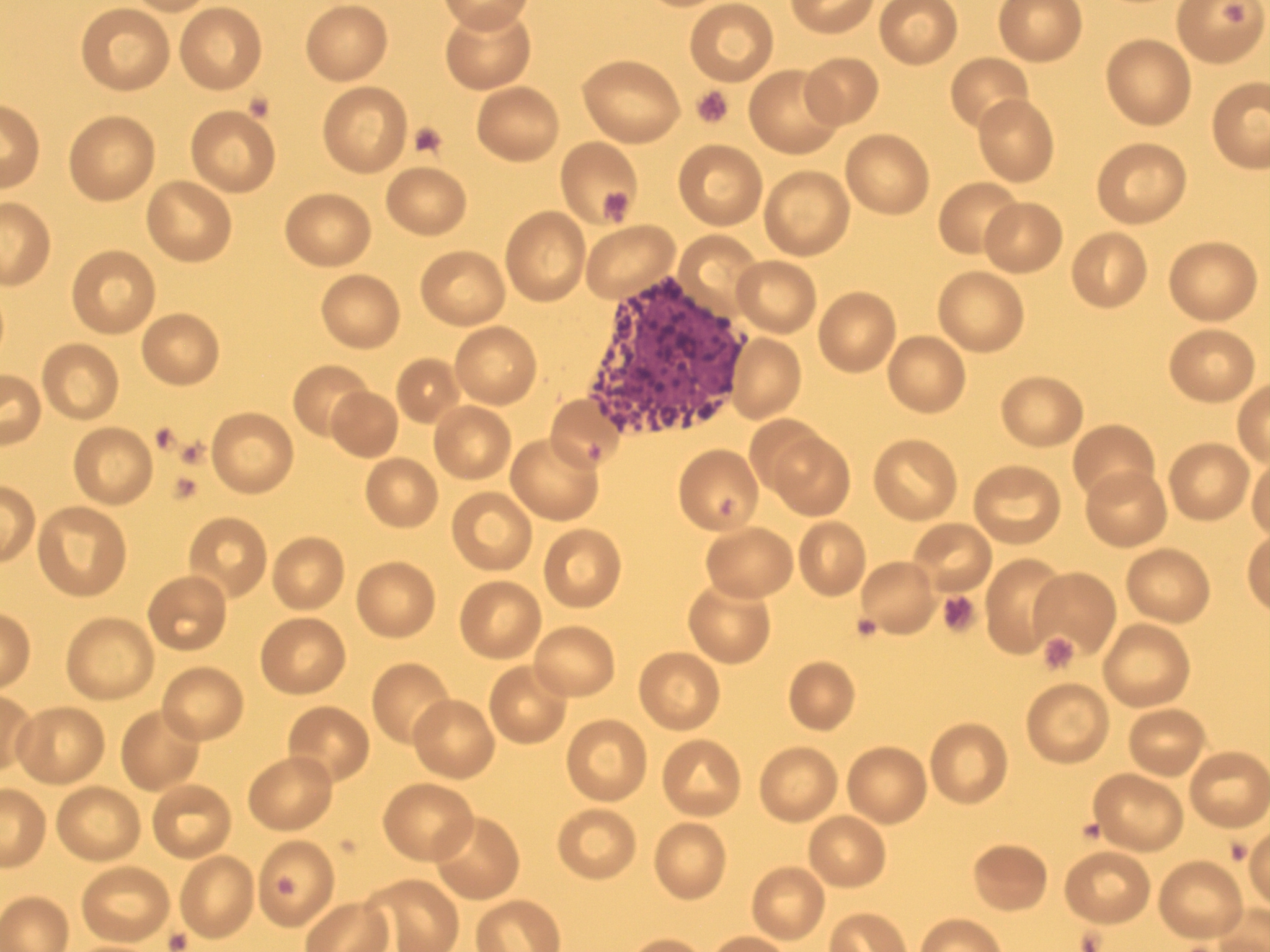
- An image from a peripheral blood smear showing a promyelocyte. From MLS Collection, University of Alberta, https://doi.org/10.7939/R3FQ9QM3R
-
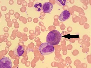
- An image from a bone marrow smear showing a promyelocyte (indicated by the arrow) and other myeloid precursors. 100x oil immersion. From MLS Collection, University of Alberta, https://doi.org/10.7939/R3QJ78D45

-
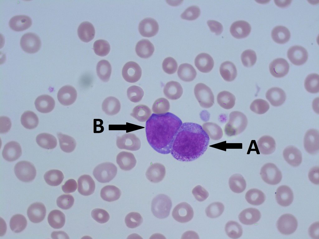
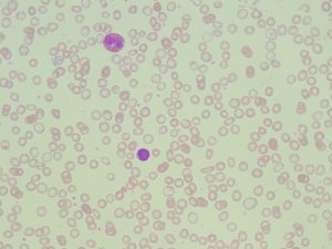
- A peripheral blood smear demonstrating a promyeloctye (A) and a blast (B). From MLS Collection, University of Alberta, https://doi.org/10.7939/R3NK36M47
Notes: Presence of primary granules marks maturation at the promyelocyte stage.3
Nucleus-to-Cytoplasm Ratio: 3:1 2
Nucleoli: 1-32
Nucleus:1-3
Round to oval
Central or eccentrically located
Reddish-blue chromatin
Fine and slightly coarser chromatin than a myeloblast
Cytoplasm:2
Lightly basophilic
Primary (fine, nonspecific) granules present (reddish-purple)
Normal % in Bone Marrow: 2-5%2
Normal % in Peripheral Blood: 0% 2
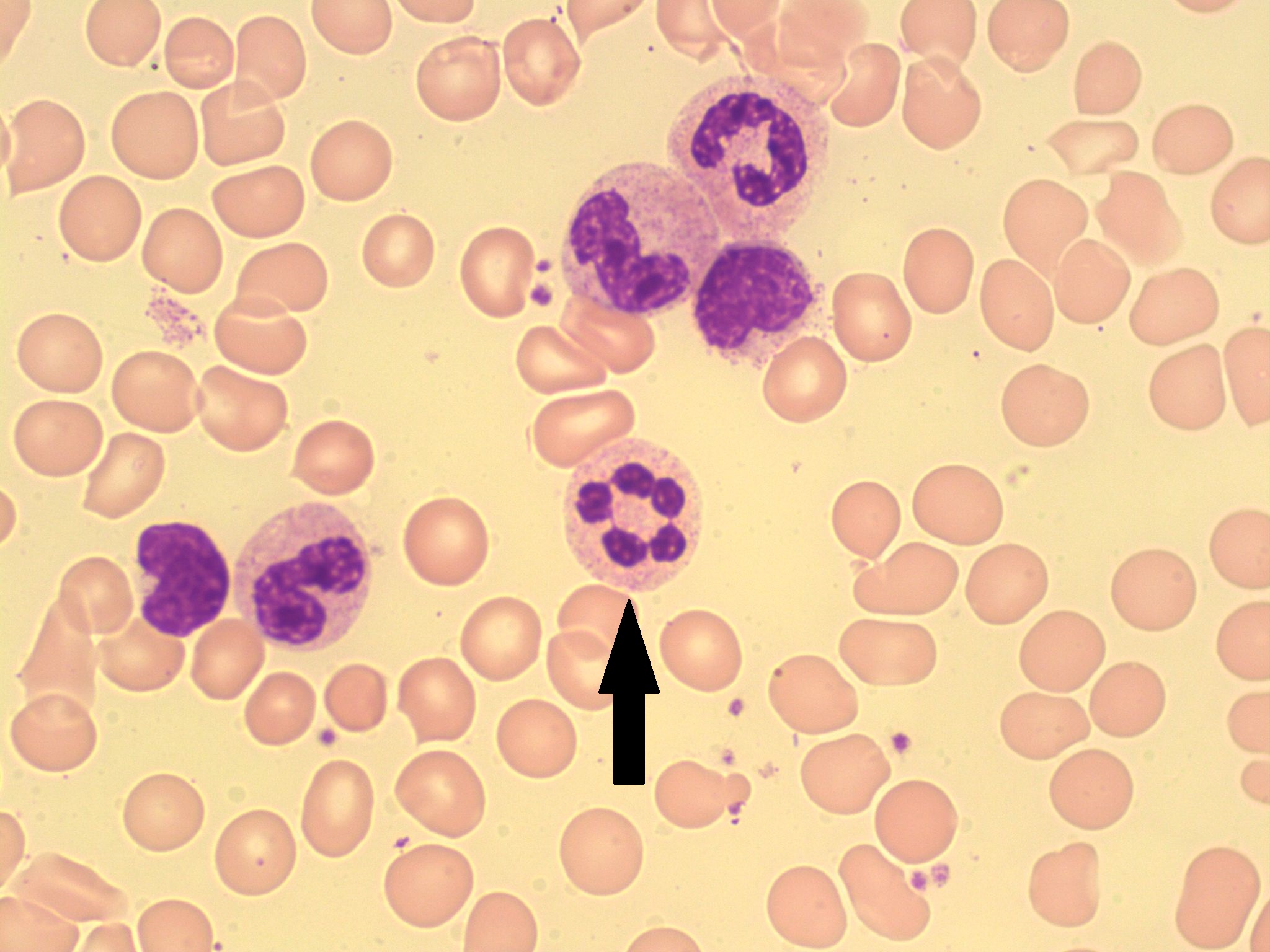
Myelocyte
-

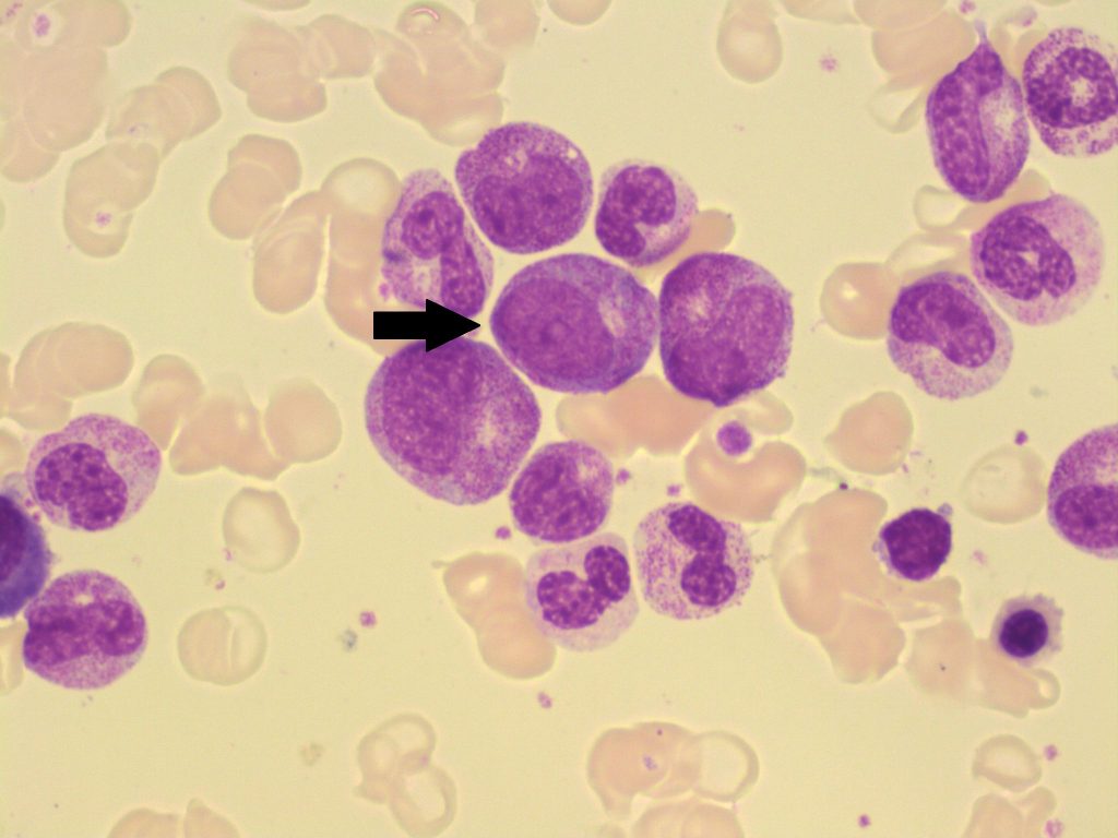
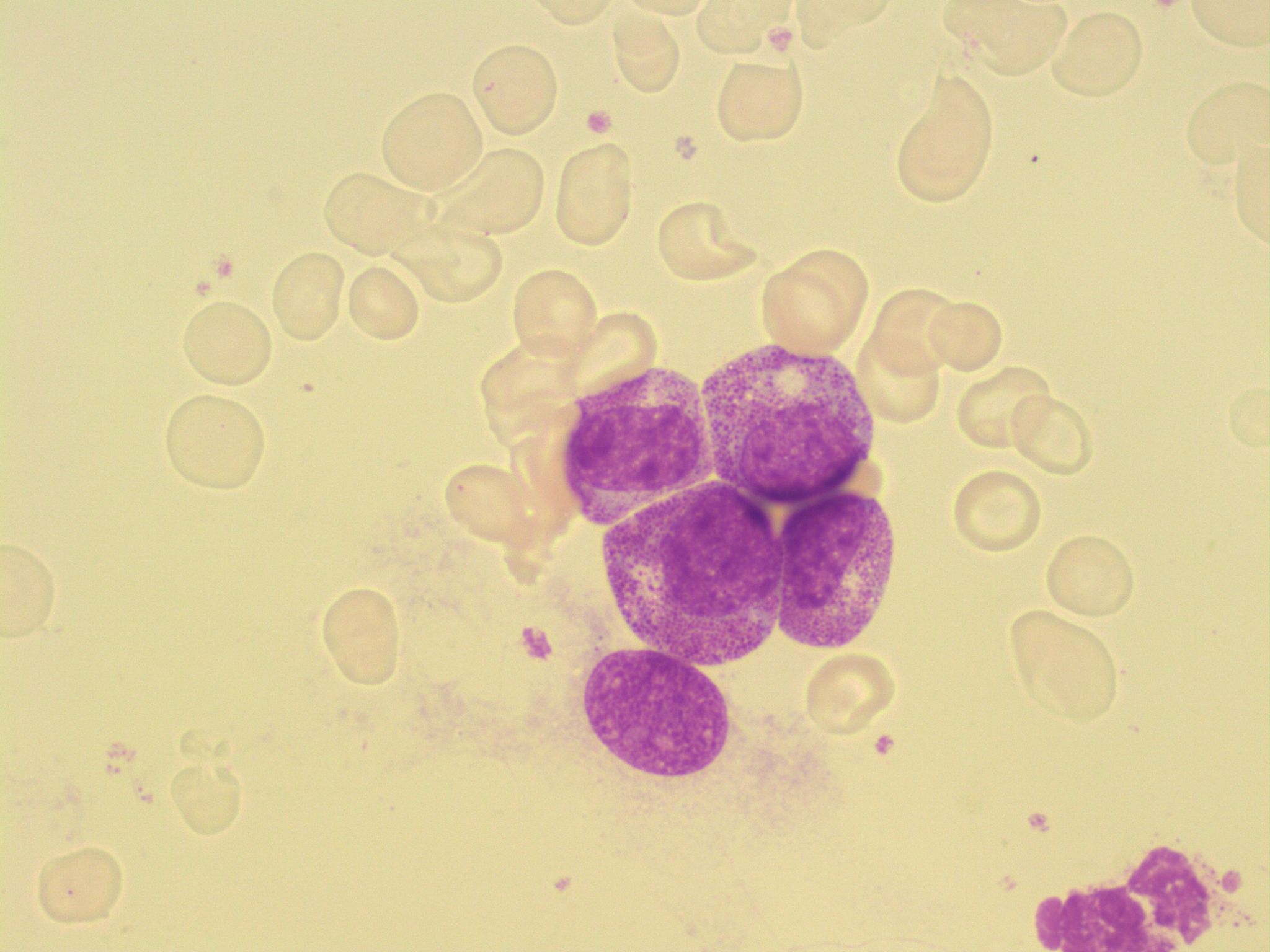
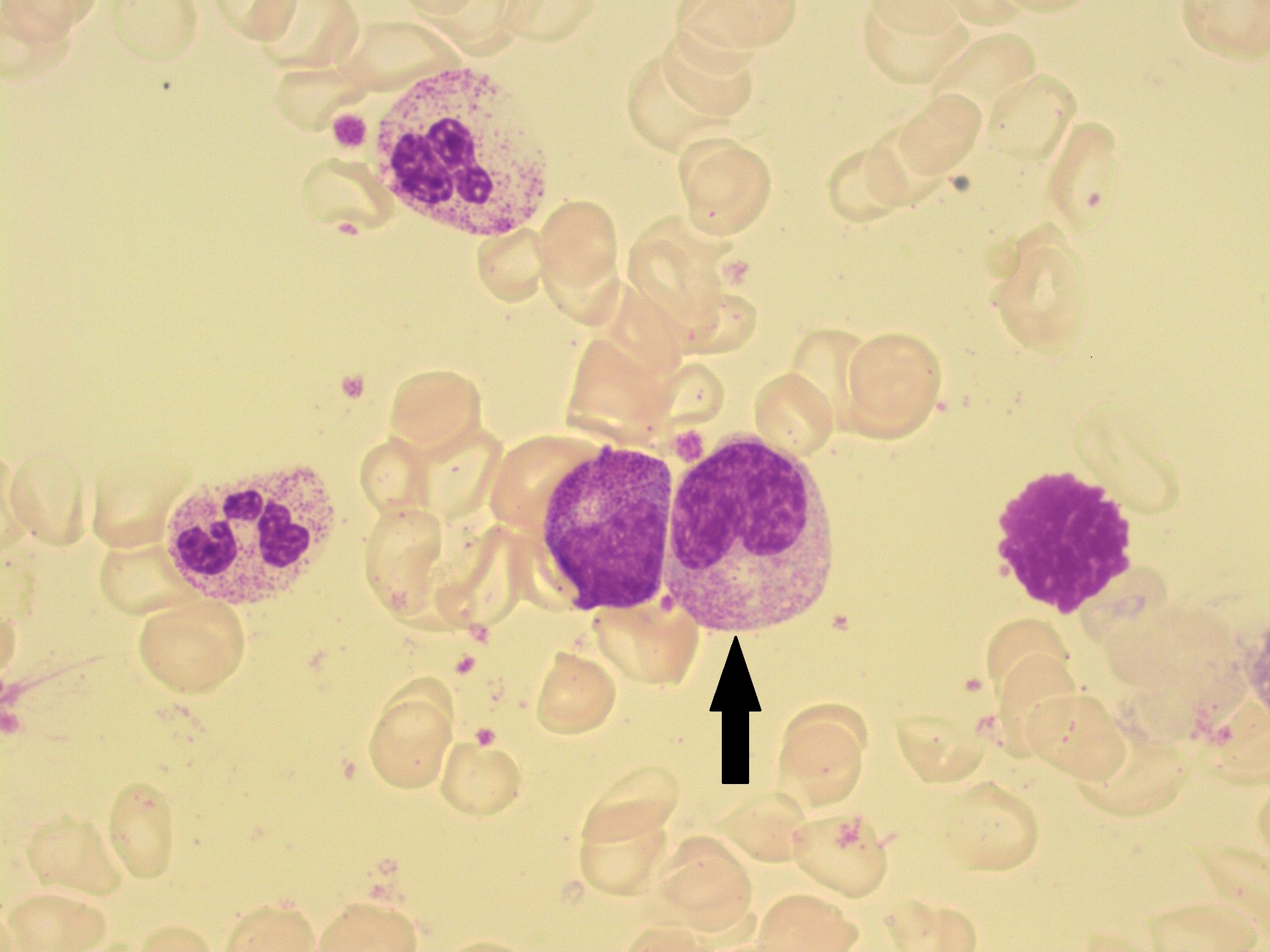
- An image from a bone marrow smear showing four myelocytes (center) with both primary and secondary granules. 100x oil immersion. From MLS Collection, University of Alberta, https://doi.org/10.7939/R3V98069S
-
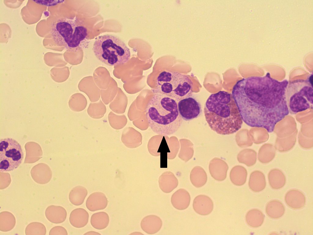
- An image from a bone marrow smearing showing a eosinophilic myelocyte (indicated by the arrow). A neutrophil myelocyte is below the eosinophil myelocyte. From MLS Collection, University of Alberta, https://doi.org/10.7939/R36M33K0X
-
- An image of a bone marrow smear with myeloid precursors. Myelocytes are indicated with arrows. 100x oil immersion. From MLS Collection, University of Alberta, https://doi.org/10.7939/R3BC3TC4P
Notes: Presence of secondary granules marks maturation at the myelocyte stage. Primary granules may still be seen but decrease in number as the cell matures. Secondary granules become more predominant as the cell mature and are considered specific to a granulocytic lineage.1
The myelocyte is the last stage where the cell is able to undergo mitosis.1
Nucleus-to-Cytoplasm Ratio: 2:1 2
Nucleoli: Usually not visible2
Nucleus:2,3
Round to oval
Eccentrically located
Reddish-purple, slightly clumped chromatin
Cytoplasm:2-5
Primary granules may be present in small amounts (Decrease in number as the cell matures).
Secondary (coarse, specific) granules present (Increase in number as the cell matures).
|
Granulocyte |
Cytoplasm Colour |
Secondary (Coarse, Specific) Granule Colour |
|
Neutrophil |
pink-tan |
azurophilic (reddish-purple) |
|
Eosinophil |
cream coloured to colourless |
eosinophilic (Pale to dark orange) |
|
Basophil |
pale blue |
basophilic (dark purple-black) |
Normal % in the Bone Marrow and Peripheral Blood:2,4,5
|
Granulocyte |
% In Bone Marrow |
% In Peripheral Blood |
|
Neutrophil |
5-19% |
0% |
|
Eosinophil |
0-2% |
0% |
|
Basophil |
0-1% |
N/A |
Metamyelocyte
-

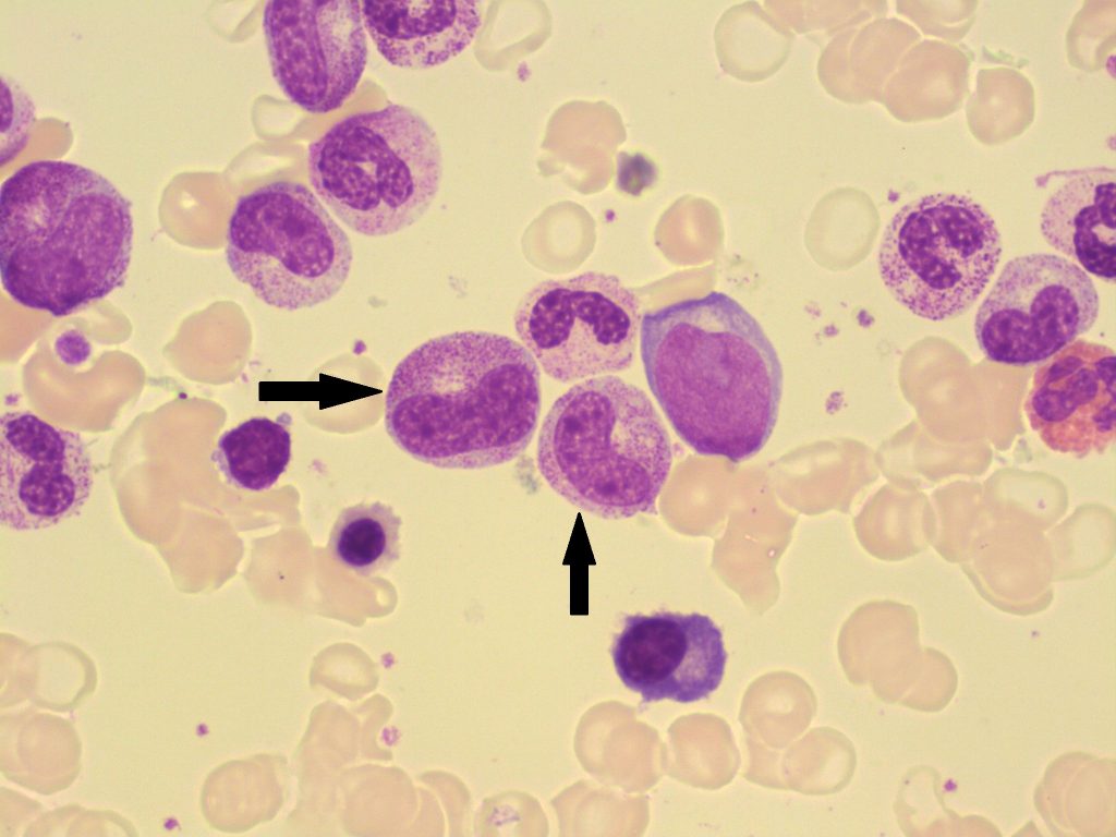
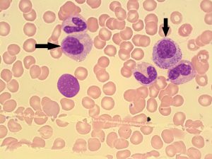
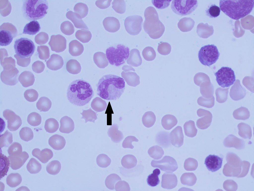
- An image from a bone marrow smear showing two metamyelocytes (indicated with arrows). 50x oil immersion. From MLS Collection, University of Alberta, https://doi.org/10.7939/R30863M9J

-

- An image from a bone marrow smear showing a metamyelocyte (indicated with an arrow). A myelocyte is present on the left of the metameylocyte. 100x oil immersion. From MLS Collection, University of Alberta, https://doi.org/10.7939/R33T9DN6V
Notes: Cell is no longer capable of mitosis at this stage. Characteristic feature of a metamyelocyte is the indented nucleus shape (nucleus looks as if it was lightly poked).1
Nucleus-to-Cytoplasm Ratio: 1.5:12
Nucleoli: Not visible2
Nucleus:1-3
Indented (kidney bean shaped); indent is less than one-third of the diameter of the hypothetical round nucleus
Eccentrically located
Dark purple, coarse, clumped chromatin
Cytoplasm:2-5
|
Granulocyte |
Cytoplasm Colour |
Secondary (Coarse, Specific) Granule Colour |
|
Neutrophil |
pink-tan |
azurophilic (reddish-purple) |
|
Eosinophil |
cream coloured to colourless |
eosinophilic (Pale to dark orange) |
|
Basophil |
pale blue |
basophilic (dark purple-black) |
Normal % in the Bone Marrow and Peripheral Blood:2-5
|
Granulocyte |
% In Bone Marrow |
% In Peripheral Blood |
|
Neutrophil |
3-22% |
0% |
|
Eosinophil |
0-2% |
0% |
|
Basophil |
0-1% |
N/A |
Band
-
- An image from a bone marrow smear demonstrating a band (indicated by an arrow) .100x oil immersion. From MLS Collection, University of Alberta, https://doi.org/10.7939/R3G44J599
-
- An image from a bone marrow smear demonstrating a band (indicated by an arrow). From MLS Collection, University of Alberta, https://doi.org/10.7939/R36Q1SZ5T

Notes: Stage shows a nucleus with a larger indentation than a metamyelocyte but it still considered non-segmented.1
Nucleus-to-Cytoplasm Ratio: Cytoplasm predominates 2
Nucleoli: Not visible2
Nucleus:1,3
Indentation takes up more than one-third of the diameter of the hypothetical round nucleus.
Appears C, U, or S shaped
Centrally or eccentrically located
Dark purple, coarse, clumped chromatin
Cytoplasm:2-5
|
Granulocyte |
Cytoplasm Colour |
Secondary (Coarse, Specific) Granule Colour |
|
Neutrophil |
pink-tan |
azurophilic (reddish-purple) |
|
Eosinophil |
cream coloured to colourless |
eosinophilic (Pale to dark orange) |
|
Basophil |
pale blue |
basophilic (dark purple-black) |
Normal % in the Bone Marrow and Peripheral Blood:2,4,5
|
Granulocyte |
% In Bone Marrow |
% In Peripheral Blood |
|
Neutrophil |
7-33% |
0-5% |
|
Eosinophil |
0-2% |
Rare |
|
Basophil |
0-1% |
N/A |
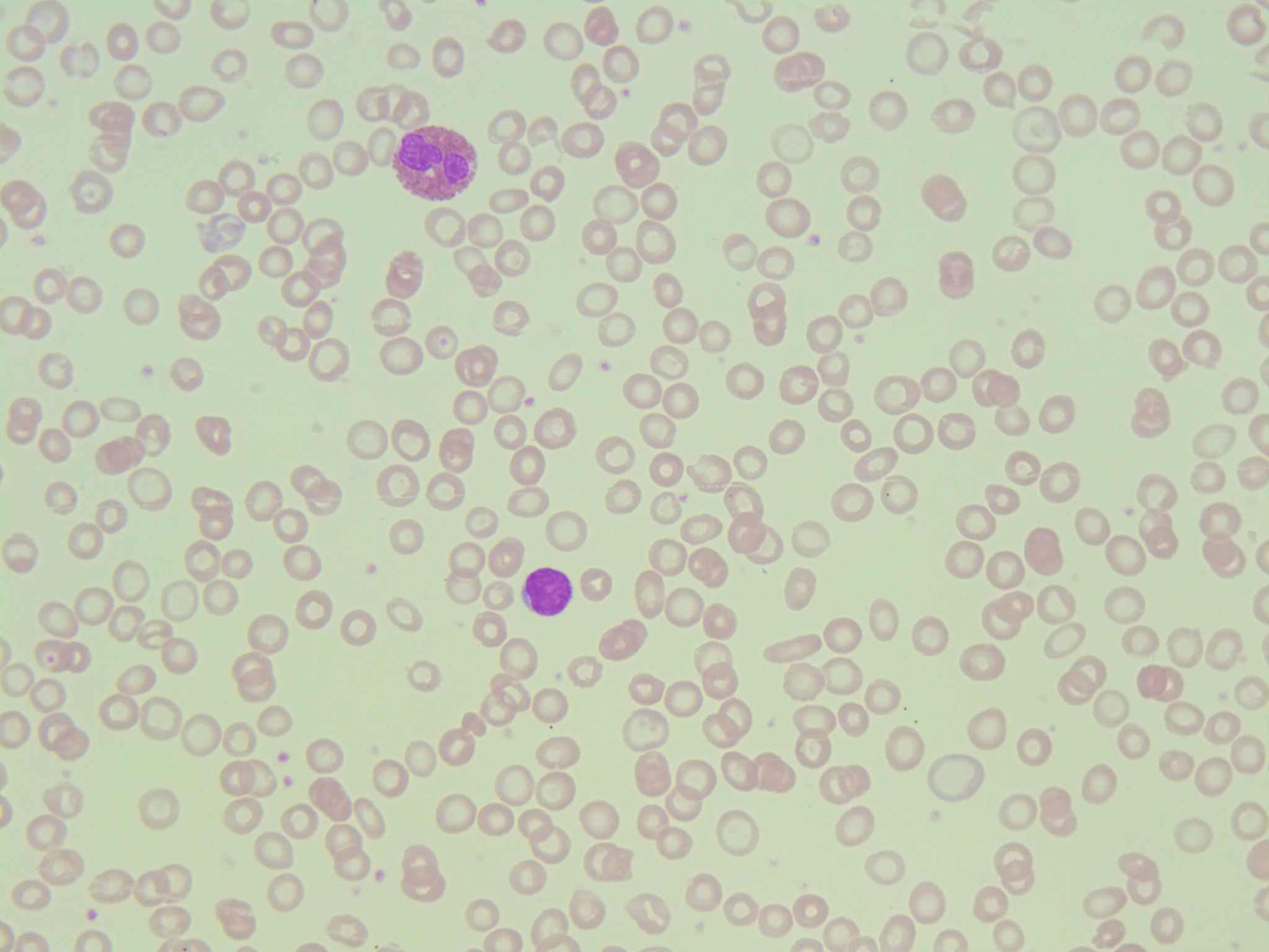
Mature (Segmented) Granulocyte
-
- An image from a bone marrow smear showing a mature, segmented neutrophil (indicated by an arrow). 100x oil immersion. From MLS Collection, University of Alberta, https://doi.org/10.7939/R31V5BV7D
-
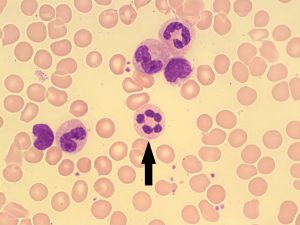
- An image from a peripheral blood smear showing a neutrophil. 100x oil immersion. From MLS Collection, University of Alberta, https://doi.org/10.7939/R39Z90T0G

-
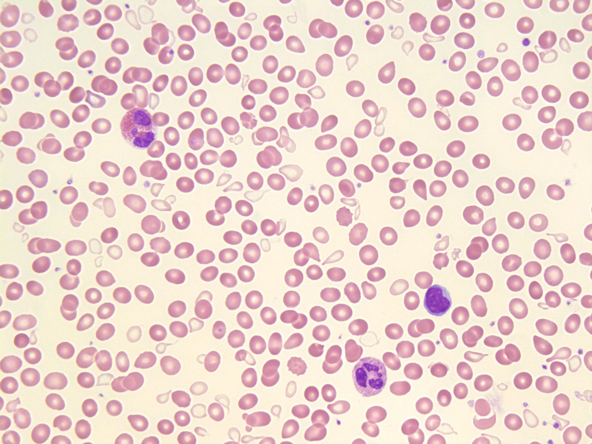
- An image from a peripheral blood smear with a mature eosinophil (top left). 50x oil immersion. From MLS Collection, University of Alberta, https://doi.org/10.7939/R3599ZH07
-
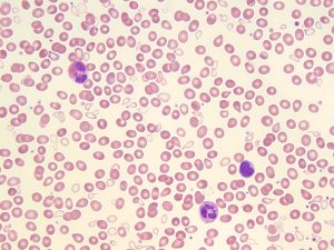
- A peripheral blood smear picture showing a mature eosinophil (top left) and neutrophil (bottom right). 50x oil immersion. From MLS Collection, University of Alberta, https://doi.org/10.7939/R3V11W18D
-
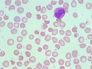
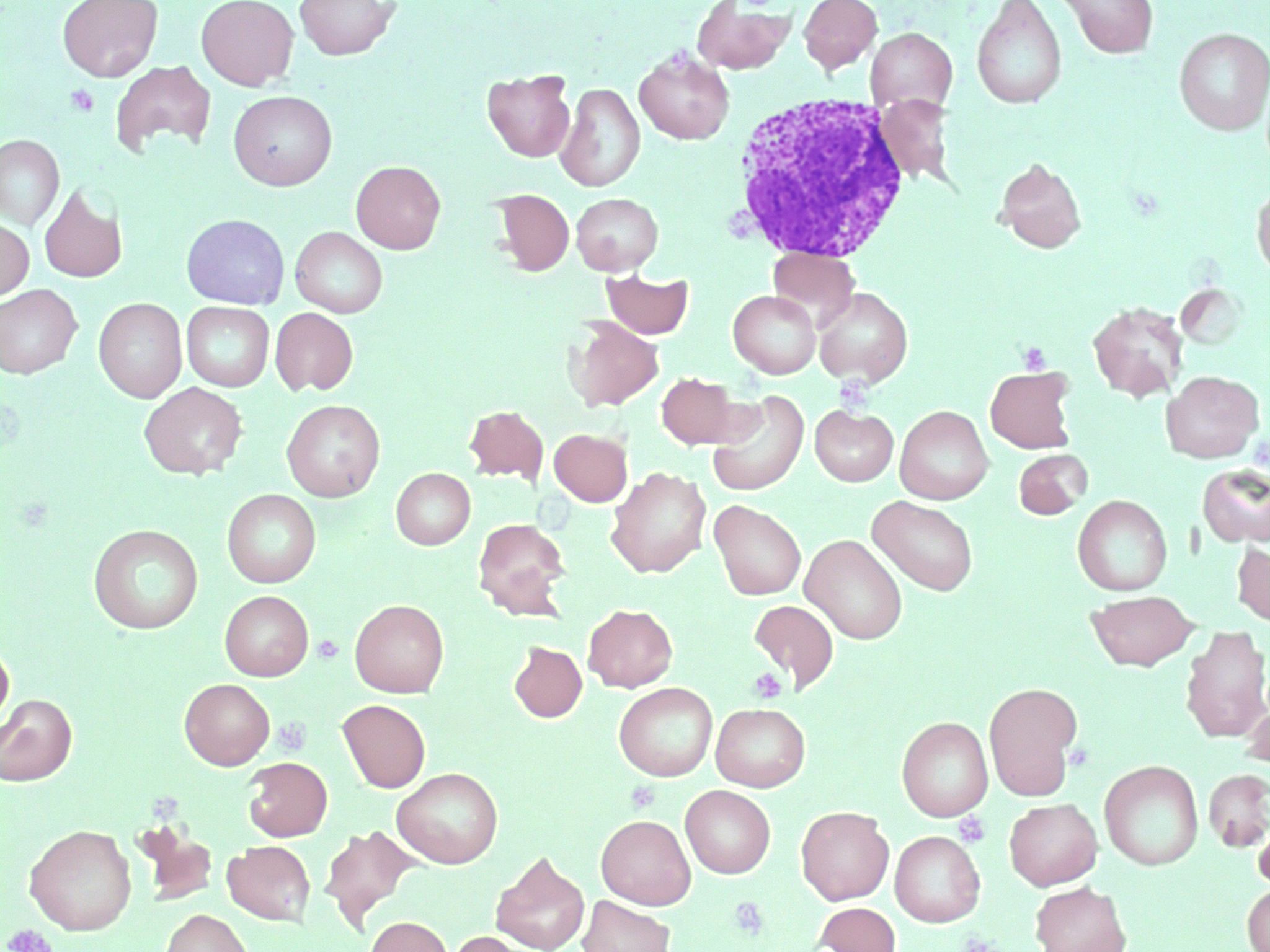
- An image from a normal peripheral blood smear showing a basophil. 100x oil immersion. From MLS Collection, University of Alberta, https://doi.org/10.7939/R30K26S7N
-
- An image from a normal peripheral blood smear showing a basophil. 100x oil immersion. From MLS Collection, University of Alberta, https://doi.org/10.7939/R34Q7R548
Nucleus-to-Cytoplasm Ratio: Cytoplasm predominates
Nucleoli: Not visible2
Nucleus:1-5
Centrally or eccentrically located
Coarse, clumpy, dark purple staining chromatin
Nucleus is separated into lobes which are all connected by chromatin filaments:
|
Granulocyte |
Normal Number of Segmented Lobes |
|
Neutrophil |
2-5 |
|
Eosinophil |
2-3 |
|
Basophiil |
Usually 2, often obscured by granules |
Cytoplasm:2,4,5
|
Granulocyte |
Cytoplasm Colour |
Secondary (Coarse, Specific) Granule Colour |
|
Neutrophil |
pink-tan |
azurophilic (reddish-purple) |
|
Eosinophil |
cream coloured to colourless |
eosinophilic (Pale to dark orange) |
|
Basophil |
pale blue |
basophilic (dark purple-black), often obscure the nucleus |
Normal % in the Bone Marrow and Peripheral Blood:2,4,5
|
Granulocyte |
% In Bone Marrow |
% In Peripheral Blood |
|
Neutrophil |
3-11% |
50-70% |
|
Eosinophil |
0-3% |
0-5% |
|
Basophil |
<1% |
0-1% |
References:
1. Landis-Piwowar K. Granulocytes and Monocytes. In: Clinical laboratory hematology. 3rd ed. New Jersey: Pearson; 2015. p. 97-121.
2. Rodak BF, Carr JH. Neutrophil maturation. In: Clinical hematology atlas. 5th ed. St. Louis, Missouri: Elsevier Inc.; 2017. p. 41-54.
3. Bell A, Harmening DM, Hughes VC. Morphology of human blood and marrow cells. In: Clinical hematology and fundamentals of hemostasis. 5th ed. Philadelphia: F.A. Davis Company; 2009. p. 1-41.
4. Rodak BF, Carr JH. Eosinophil maturation. In: Clinical hematology atlas. 5th ed. St. Louis, Missouri: Elsevier Inc.; 2017. p. 65-74.
5. Rodak BF, Carr JH. Basophil maturation. In: Clinical hematology atlas. 5th ed. St. Louis, Missouri: Elsevier Inc.; 2017. p. 75-8.
