
66 Chediak-Higashi Syndrome
Michelle To and Valentin Villatoro
-
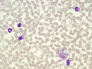
- An image of a peripheral blood smear demonstrating neutrophils with abnormally large fused granules seen in Chediak-Higashi Syndrome. 50x oil immersion. From MLS Collection, University of Alberta, https://doi.org/10.7939/R39S1M158
-
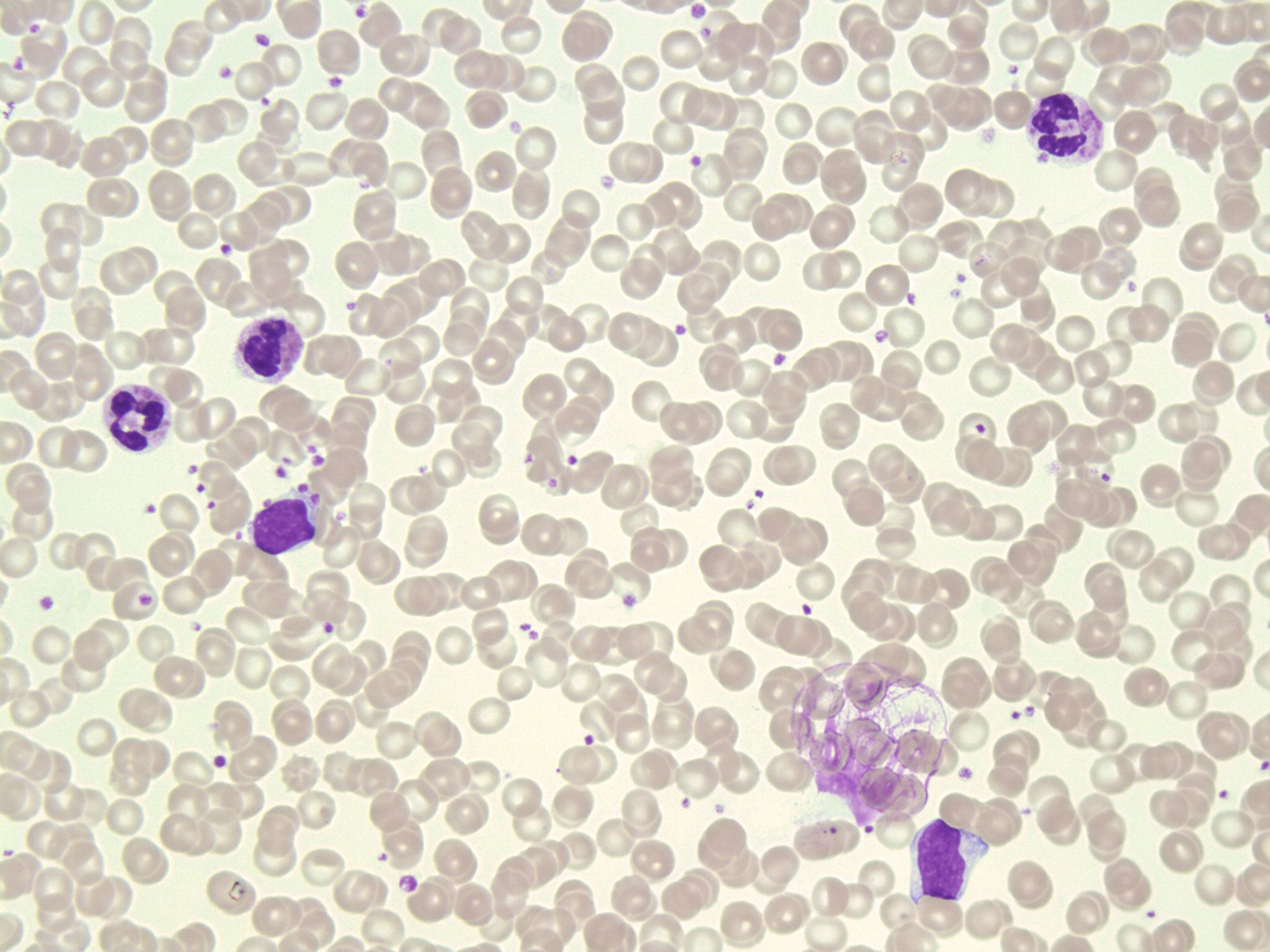
- An image of a peripheral blood smear demonstrating a neutrophil with abnormally large fused granules (top) and a lymphocyte containing a single large granule in the cytoplasm (bottom) seen in Chediak-Higashi syndrome. 50x oil immersion. From MLS Collection, University of Alberta, https://doi.org/10.7939/R3707X414
-

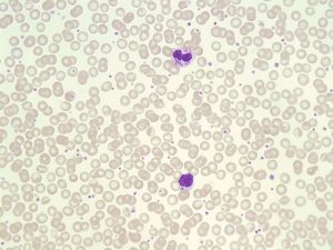
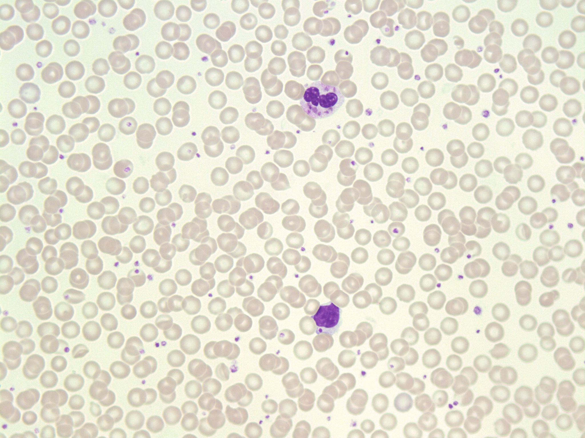
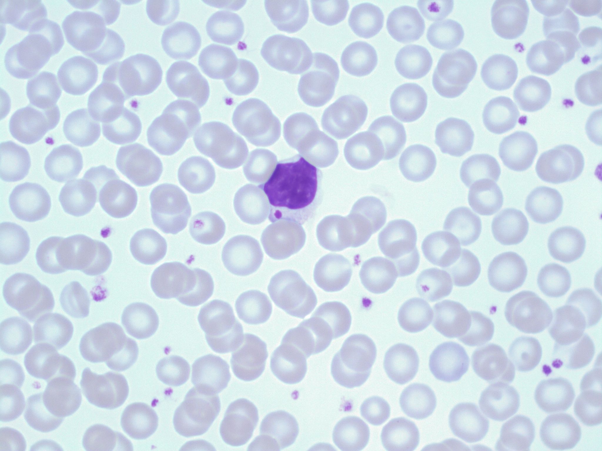
- An image of a peripheral blood smear demonstrating a lymphocyte containing a single large granule in the cytoplasm seen in Chediak-Higashi syndrome. 100X oil immersion. From MLS Collection, University of Alberta, https://doi.org/10.7939/R3377697R
PBS Key Features:1,2
Leukocytes contain abnormally large lysosomal granules in the cytoplasm. Granules represent the aggregation of primary granules combined with the fusion of secondary granules.
Cause:3
Mutation in the CHS1/LYST gene which encodes for a vesicle transport protein.
Inheritance Pattern:3-5
Autosomal recessive
Clinical Significance:1,3-5
Development of lysosomes are abnormal resulting in the fusion of granules. The syndrome results in impaired chemotaxis, defective degranulation, and defective killing of bacteria. Granulocytes, Platelets, Monocytes, and lymphocytes are dysfunctional.
Patients often present with oculocutaneous albinism, recurrent bacterial infections and bleeding tendencies. Complications develop during early childhood.
CBC:1,2,5
Anemia
Neutropenia
Thrombocytopenia
References:
1. Harmening DM, Marty J, Strauss RG. Cell biology, disorders of neutrophils, infectious mononucleosis, and reactive lymphocytosis. In: Clinical hematology and fundamentals of hemostasis. 5th ed. Philadelphia: F.A. Davis Company; 2009. p. 305-30.
2. Bain BJ. Morphology of blood cells. In: Blood cells: a practical guide [Internet]. 5th ed. Chichester, UK: John Wiley & Sons, Ltd; 2015 [cited 2018 Jul 10]: 67-185. Available from: http://doi.wiley.com/10.1002/9781118817322
3. Marionneaux S. Nonmalignant leukocyte disorders. In: Rodak’s hematology clinical applications and principles. 5th ed. St. Louis, Missouri: Saunders; 2015. p. 475-97.
4. Turgeon ML. Nonmalignant Disorders of Granulocytes and monocytes. In: Clinical hematology: theory and procedures. 4th ed. Philadelphia, PA: Lippincott Williams & Wilkins; 1999. p. 206-16.
5. Landis-Piwowar K. Nonmalignant disorders of leukocytes: granulocytes and monocytes. In: Clinical laboratory hematology. 3rd ed. New Jersey: Pearson; 2015. p. 388-407.
