
6 Bite (Keratocyte) & Blister (Helmet) Cells
Michelle To and Valentin Villatoro
-
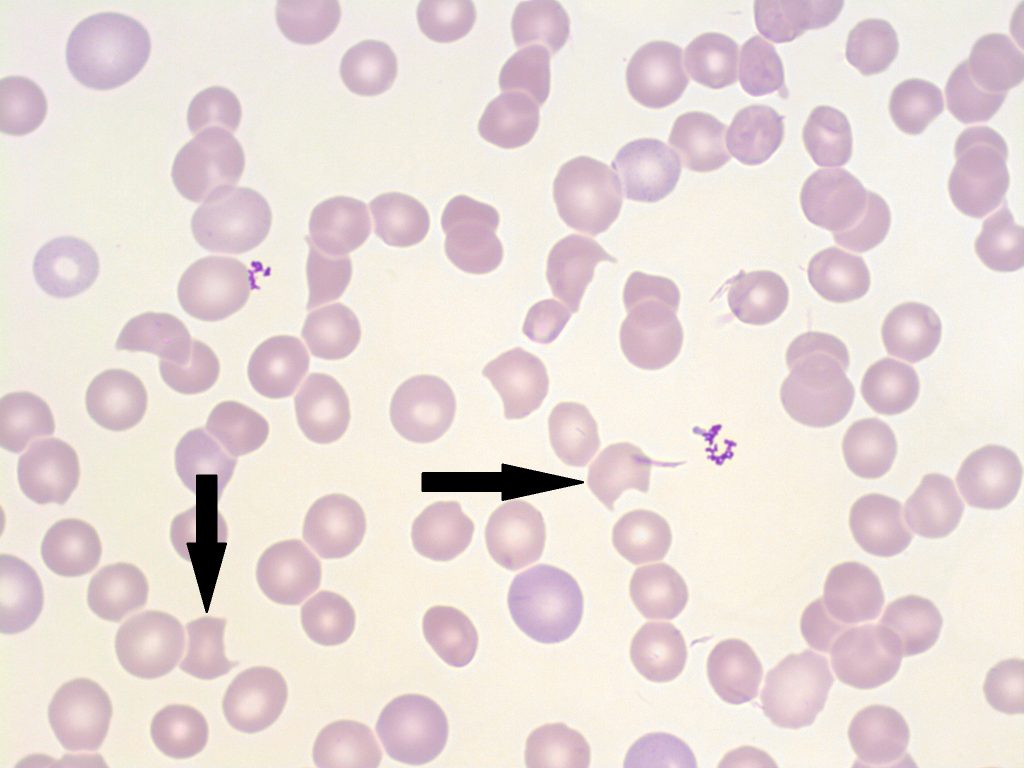
- An image of a peripheral blood smear with bite cells present (indicated with arrows). 100x oil immersion. From MLS Collection, University of Alberta, https://doi.org/10.7939/R3CC0V83F
-
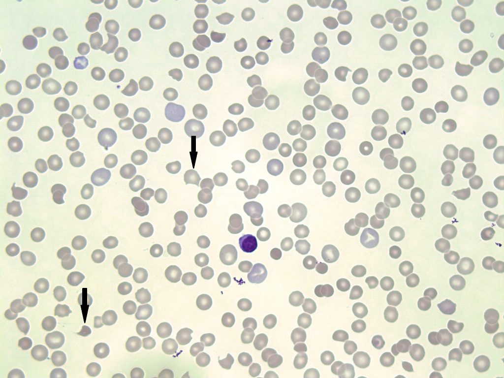
- An image of a peripheral blood smear with bite cells present (indicated with arrows). 100x oil immersion. From MLS Collection, University of Alberta, https://doi.org/10.7939/R3V698T3M

-

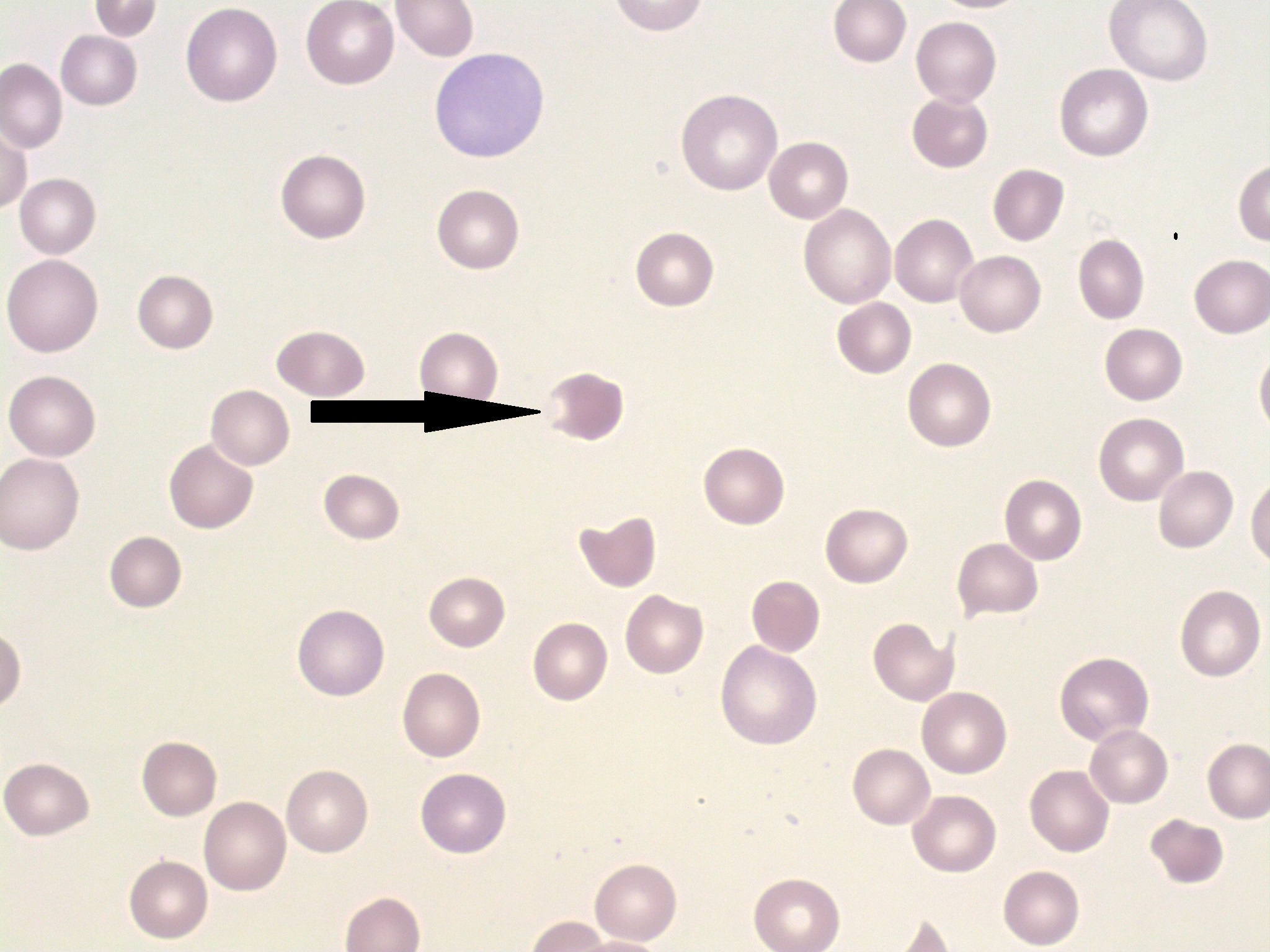
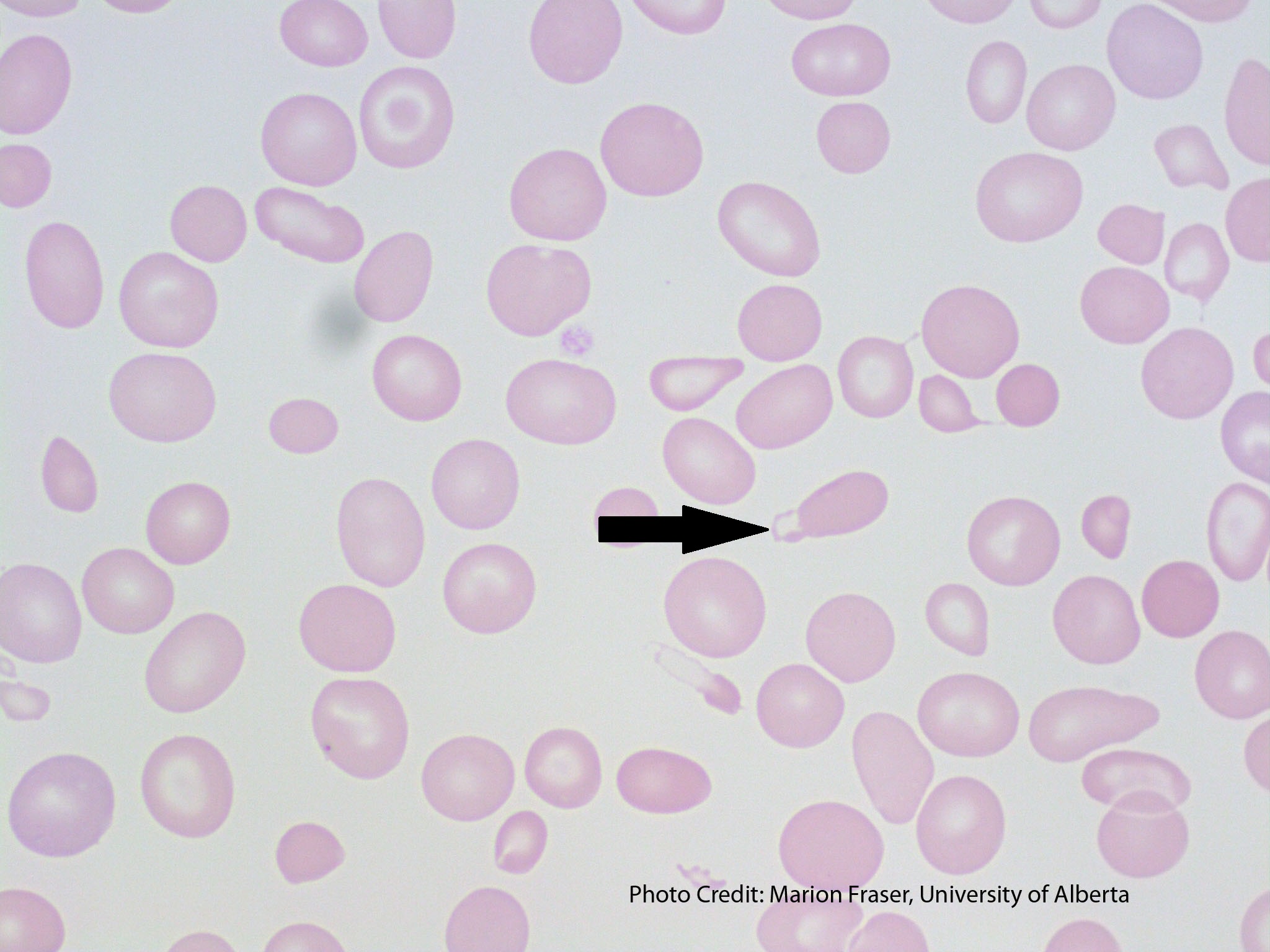
- A peripheral blood smear demonstrating a blister cell (shown with an arrow). 100x oil immersion. From MLS Collection, University of Alberta, https://doi.org/10.7939/R3H12VP5B
-
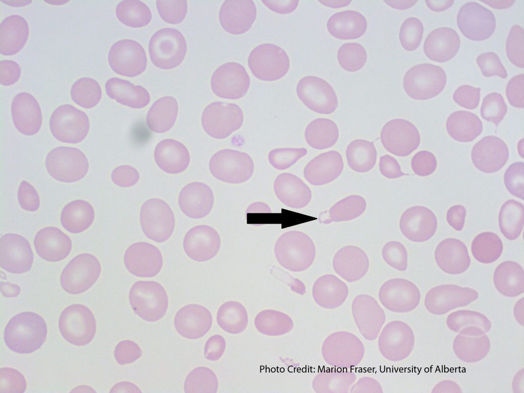
- A peripheral blood smear with a blister cell present (shown with an arrow). From MLS Collection, University of Alberta, https://doi.org/10.7939/R3HQ3SD75
Cell Description:
Bite cells are red blood cells that contain a semi-circular indent on the edge of their membrane, giving the appearance of a bite being taken out of the cell.1 Blister cells on the other hand, have cytoplasmic projections that fuse together, creating a vacuole on the edge of the membrane, giving the appearance of a blister.2
Cell Formation:
Bite and Blister cells are often seen together, and may form through various mechanisms. Red blood cells originally containing inclusions are “pitted” or removed by macrophages in the spleen, resulting in bite or blister cells.3 When the red blood cell is impaled by fibrin strands, the membrane can reform and produce a vacuole which results in a blister cell.2,3
Bite cells can also form when a blister cell ruptures.4
Associated Disease/Clinical States:2,4,5
Microangiopathic Hemolytic Anemias (MAHAs)
Mechanical Hemolysis (i.e. mechanical heart-valves)
Heinz body hemolytic anemias (G6PD Deficiency, Thalassemia)
Note: Bite and blister cells are mainly seen in clinical states where Heinz bodies are formed.2
References:
1. Ford J. Red blood cell morphology. Int J Lab Hematol [Internet]. 2013 Mar 9 [cited 2018 Jul 12];35(3):351–7. Available from: https://doi.org/10.1111/ijlh.12082
2. Landis-Piwowar K, Landis J, Keila P. The complete blood count and peripheral blood smear evaluation. In: Clinical laboratory hematology. 3rd ed. New Jersey: Pearson; 2015. p. 154-77.
3. Jones KW. Evaluation of cell morphology and introduction to platelet and white blood cell morphology. In: Clinical hematology and fundamentals of hemostasis. 5th ed. Philadelphia: F.A. Davis Company; 2009. p. 93-116.
4. Turgeon ML. Erythrocyte morphology and inclusions. In: Clinical hematology: theory and procedures. 4th ed. Philadelphia, PA: Lippincott Williams & Wilkins; 1999. p. 99-111.
5. Julius CJ, Schaub CR. Hypoproliferative anemia: anemia associated with systemic diseases. In: Clinical hematology and fundamentals of hemostasis. 5th ed. Philadelphia: F.A. Davis Company; 2009. p. 280-304
