
25 Pappenheimer Bodies (Siderotic Granules)
Michelle To and Valentin Villatoro
-
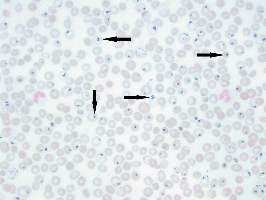
- An iron stained peripheral blood smear with pappenheimer bodies present (indicated with arrows). Perls Prussian Blue. 50x oil immersion. From MLS Collection, University of Alberta, https://doi.org/10.7939/R3FN1173T

-
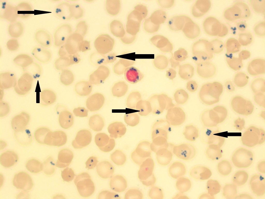
- An iron stained peripheral blood smear with pappenheimer bodies present (indicated with arrows). Perls Prussian Blue. 50x oil immersion. From MLS Collection, University of Alberta, https://doi.org/10.7939/R36689100
-

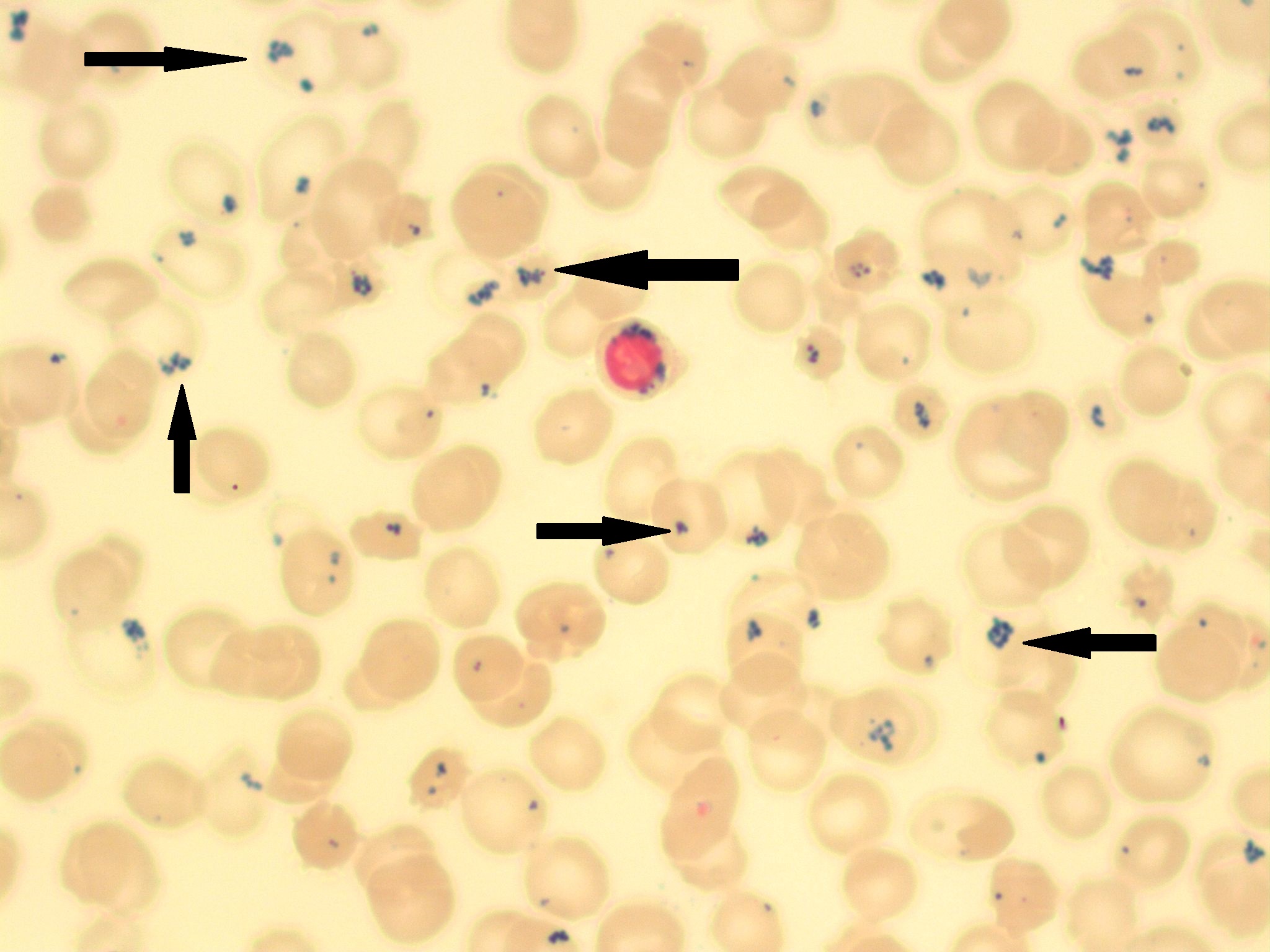
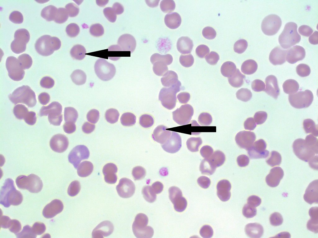
- A peripheral blood smear with pappenheimer bodies present (indicated with arrows). 100x oil immersion. From MLS Collection, University of Alberta, https://doi.org/10.7939/R35X25V0R
-
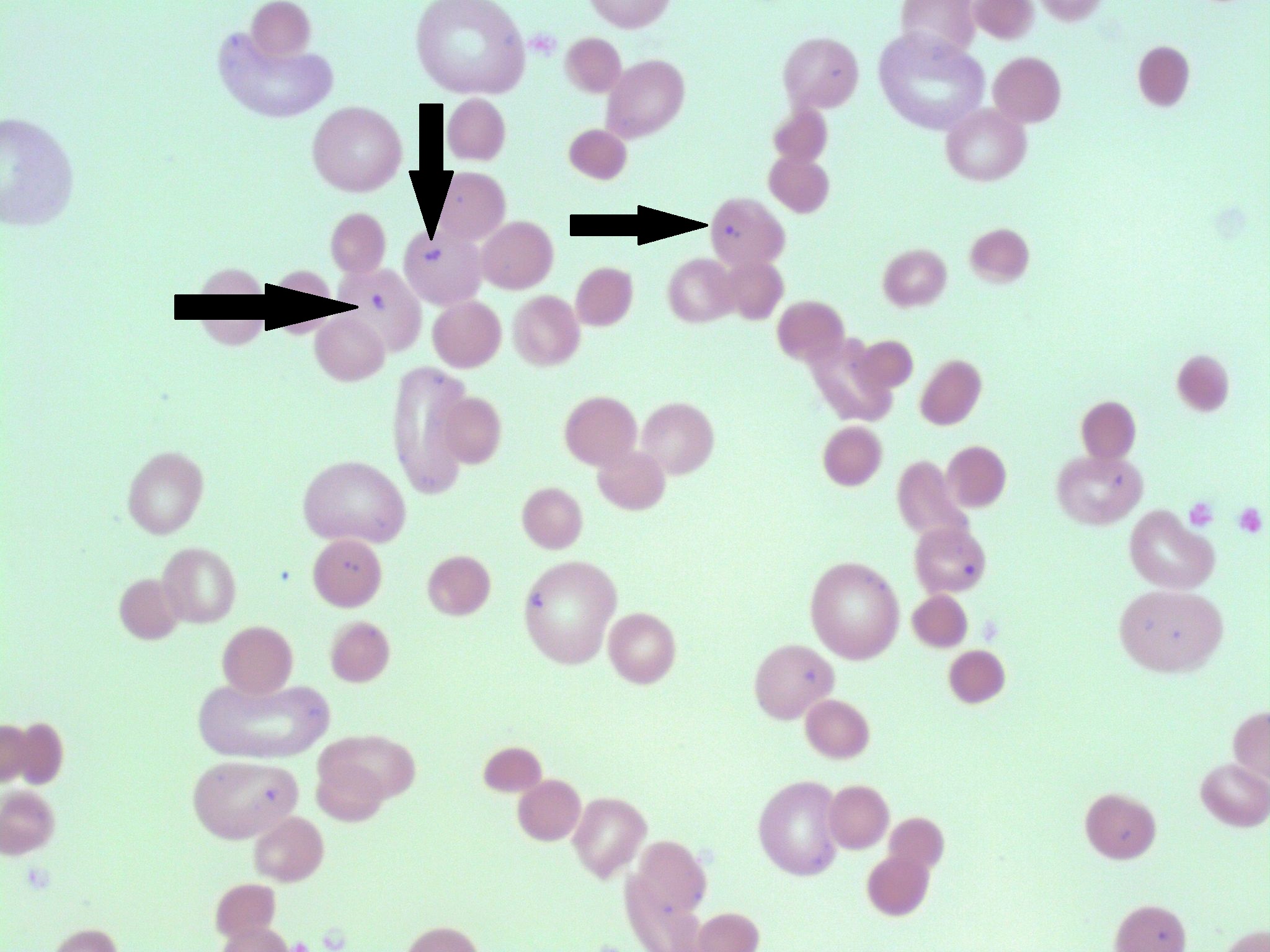
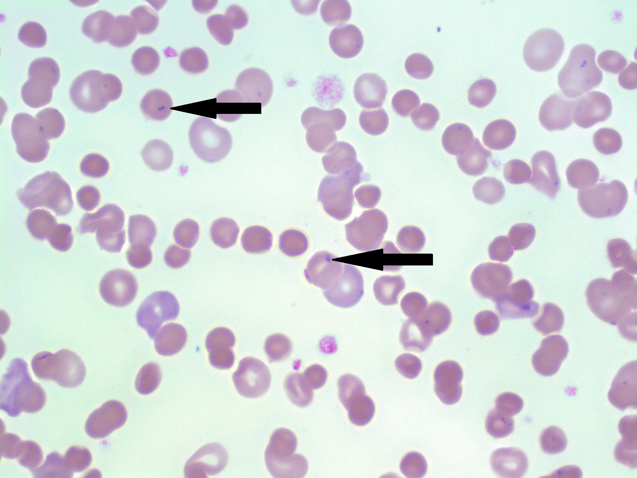
- A peripheral blood smear with pappenheimer bodies present (indicated with arrows). 100x oil immersion. From MLS Collection, University of Alberta, https://doi.org/10.7939/R3251G16Q
Appearance:
Inclusions are visible under both Wright/Romanowsky stains and Perls Prussian Blue stain. Pappenheimer inclusions appear as clusters of fine and irregular granules located at the periphery of the red blood cell.1-3
Inclusion composition:3
Iron
Associated Disease/Clinical States:1,2
Splenectomy
Sideroblastic Anemia
Thalassemia
Sickle Cell Disease
Hemachromatosis
References:
1. Landis-Piwowar K, Landis J, Keila P. The complete blood count and peripheral blood smear evaluation. In: Clinical laboratory hematology. 3rd ed. New Jersey: Pearson; 2015. p. 154-77.
2. Jones KW. Evaluation of cell morphology and introduction to platelet and white blood cell morphology. In: Clinical hematology and fundamentals of hemostasis. 5th ed. Philadelphia: F.A. Davis Company; 2009. p. 93-116.
3. Rodak BF, Carr JH. Inclusions in erythrocytes. In: Clinical hematology atlas. 5th ed. St. Louis, Missouri: Elsevier Inc.; 2017. p. 107-14.
