
24 Howell-Jolly Bodies
Michelle To and Valentin Villatoro
-
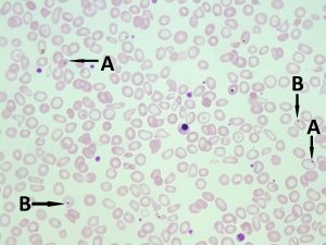
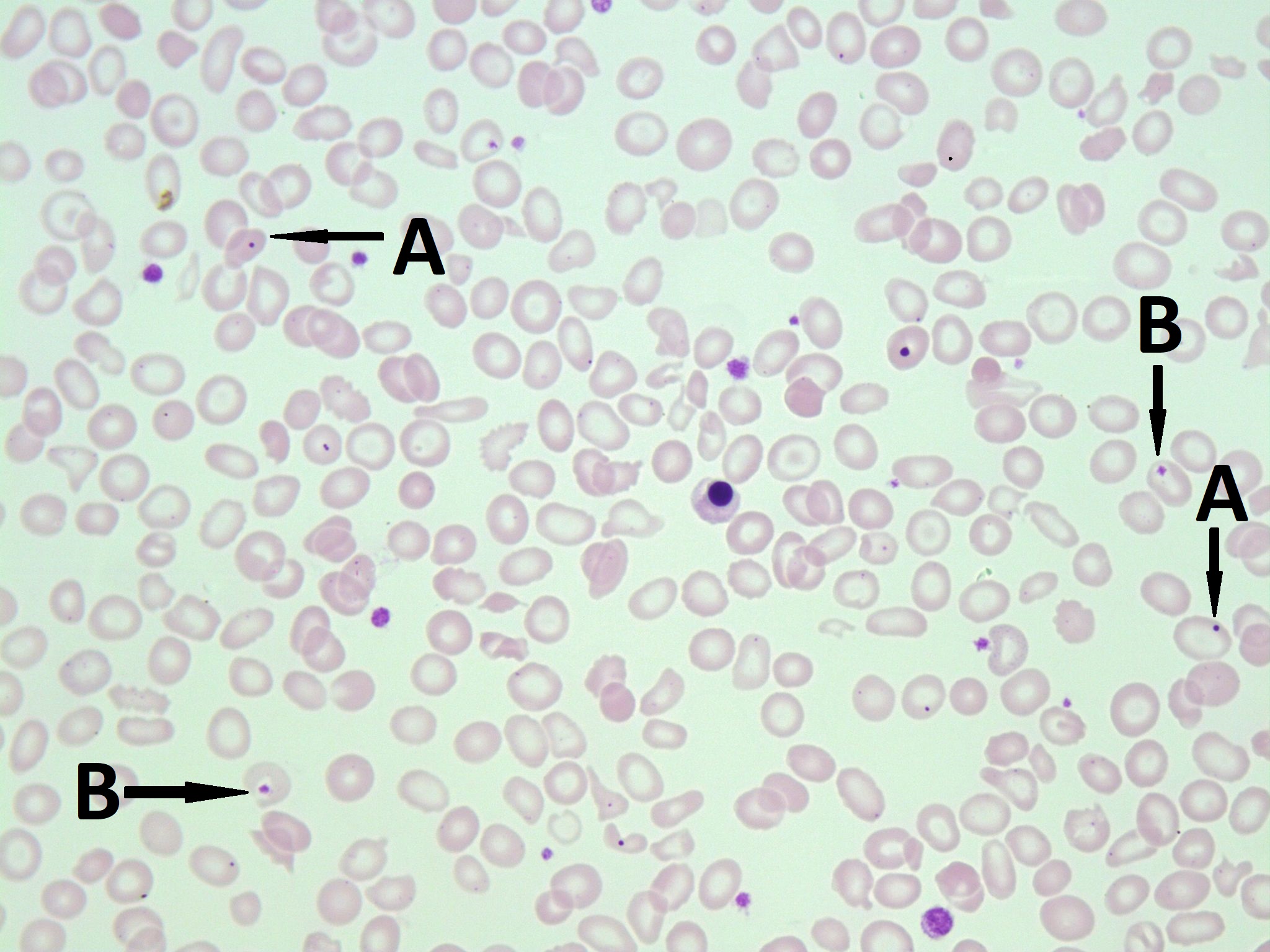
- A peripheral blood smear with Howell-Jolly bodies. A. shows Howell-Jolly bodies. B. shows platelets on top of a red blood cell. Note the clear space surrounding the platelet. 50x oil immersion. From MLS Collection, University of Alberta, https://doi.org/10.7939/R30R9MK3C
-
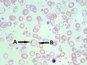
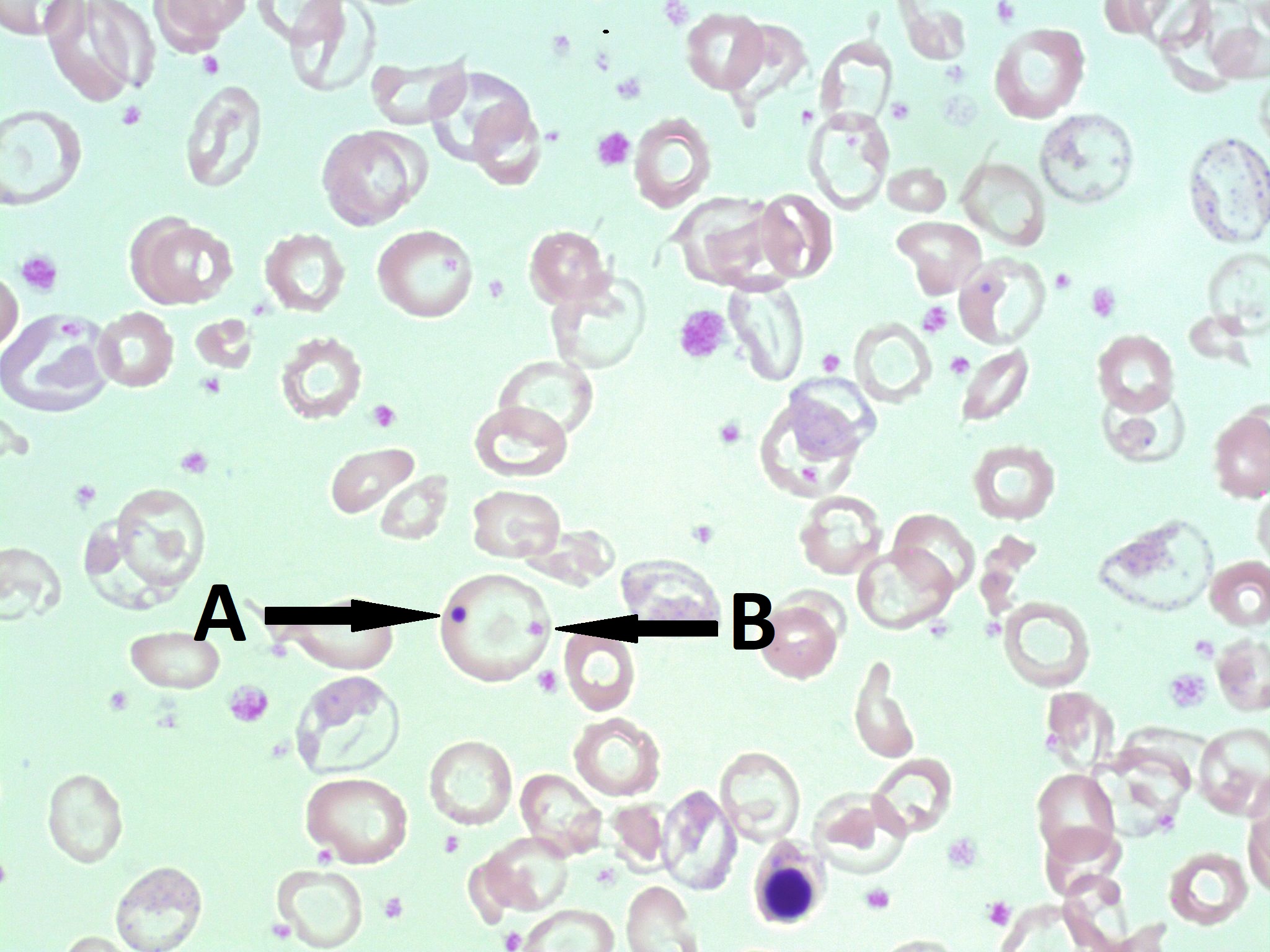
- A peripheral blood smear with a Howell-Jolly body and a platelet on the same red blood cell. A. shows Howell-Jolly body. B. shows platelets on top of a red blood cell. 100x oil immersion. From MLS Collection, University of Alberta, https://doi.org/10.7939/R3B853Z9R
-

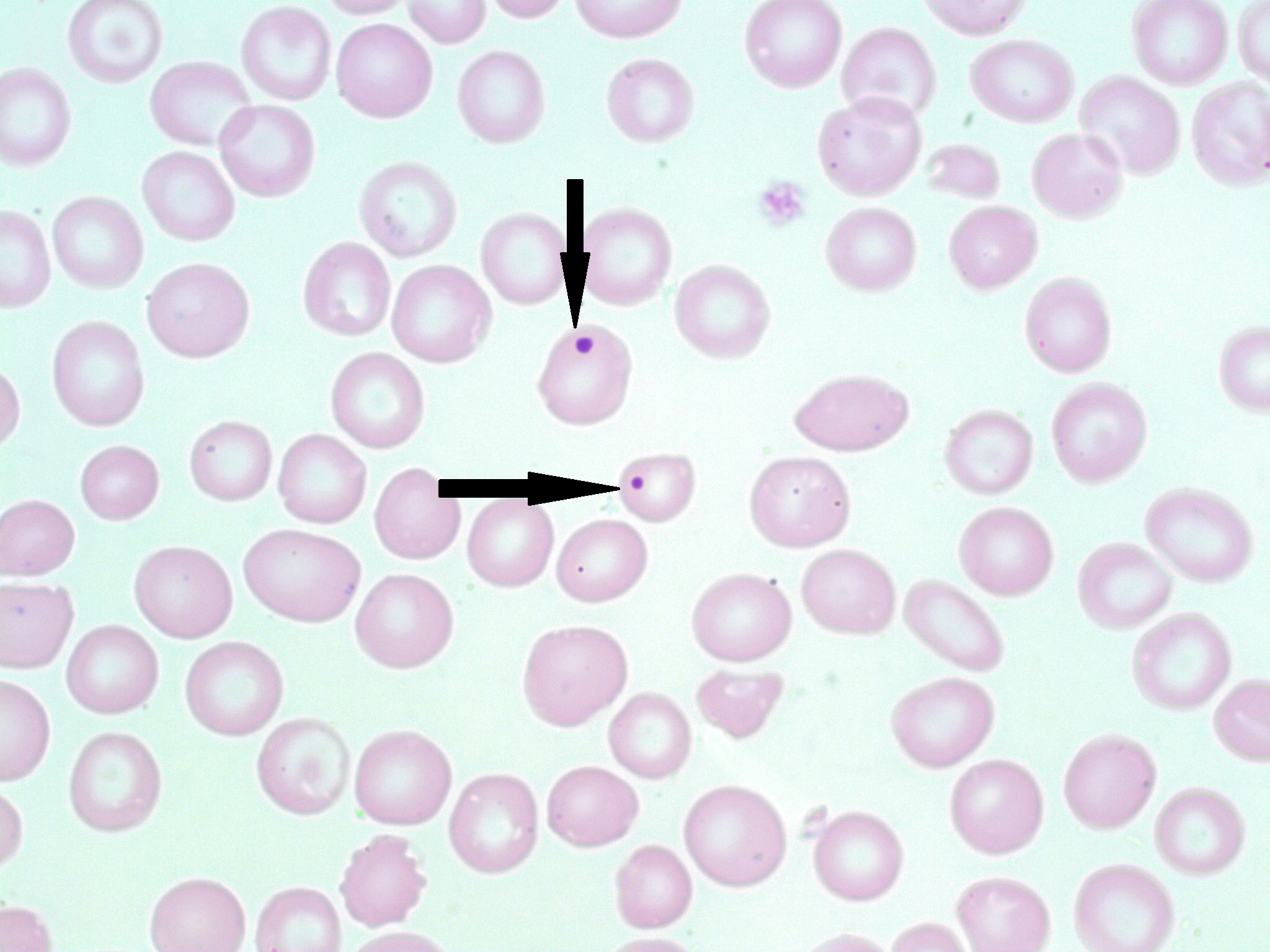
- A peripheral blood smear with Howell-Jolly bodies (indicated with arrows). 100x oil immersion. From MLS Collection, University of Alberta, https://doi.org/10.7939/R3028PV21
Appearance:
Under Wright/Romanowksy stains, Howell-Jolly Bodies appear as dark blue/purple round inclusions located at the periphery of the RBC. They usually present as a single inclusion inside the cell. Howell-Jolly Bodies are also visible under supravital stains.1-4
Inclusion composition:2,3
Nuclear fragments/remnants made up of DNA 1-4
Associated Disease/Clinical States:
Thalassemia
Megaloblastic Anemia
Myelodysplastic Syndrome
Post-splenectomy
References:
1. Landis-Piwowar K, Landis J, Keila P. The complete blood count and peripheral blood smear evaluation. In: Clinical laboratory hematology. 3rd ed. New Jersey: Pearson; 2015. p. 154-77.
2. Jones KW. Evaluation of cell morphology and introduction to platelet and white blood cell morphology. In: Clinical hematology and fundamentals of hemostasis. 5th ed. Philadelphia: F.A. Davis Company; 2009. p. 93-116.
3. Fritsma GA. Bone marrow examination. In: Rodak’s hematology clinical applications and principles. 5th ed. St. Louis, Missouri: Saunders; 2015. p. 253-68.
4. Ford J. Red blood cell morphology. Int J Lab Hematol [Internet]. 2013 Mar 9 [cited 2018 Jul 12];35(3):351–7. Available from: https://doi.org/10.1111/ijlh.12082
