6.3 Respiratory System
Overview and Functions
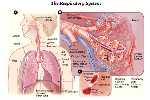
The primary functions of the respiratory system (Fig. 6.6) are to provide oxygen to the body’s tissues for cellular respiration, remove the waste product carbon dioxide, and help maintain the acid-base balance. Although cells require oxygen, it is actually the accumulation of carbon dioxide that drives the respiratory system to breathe. This system includes the muscles used to move air in and out of the lungs, the structures involved in the movement of oxygen and carbon dioxide, and the microscopic gas exchange that occurs within the lungs. The majority of the chronic pathologies that affect this system are conditions that impair the gas exchange process and result in laboured breathing and other difficulties.

(CrashCourse, 2015)
Components of the Respiratory System
Nose: The nose is the main entrance and exit for the respiratory system.
Pharynx: The pharynx is a tube made up of skeletal muscle and lined with mucous membrane that begins in the nasal cavity and ends at the larynx. The pharynx is divided into three major regions: nasopharynx, oropharynx, and laryngopharynx.
Larynx: This cartilaginous structure is found below the pharynx and connects at the lower end to the trachea. The larynx helps regulate the volume of air that enters and leaves the lungs. It is composed of three large cartilage pieces: thyroid cartilage, epiglottis, and cricoid cartilage.
Epiglottis: This very flexible, elastic piece of cartilage covers the opening of the trachea and is attached to the thyroid cartilage. It is an important structure in that it prevents food and liquids from entering the trachea.
Trachea: Also known as the windpipe, the trachea extends from the larynx to the lungs. It branches at the end into the right and left bronchi.
Bronchi: The bronchi lead to tree-like structures in both the right and left lungs that become the smaller bronchioles and finally end in the alveolar sacs and alveoli.
Alveoli: These are the small, almost grape-like structures at the end of the alveolar ducts. This is where gas exchange occurs.
Lungs: The lungs are the major organ in the respiratory system and contain the bronchi, bronchioles, and alveoli. The main function of the lungs is to exchange oxygen and carbon dioxide.
Pleura: This serous membrane lines the thoracic cavity and surrounds the lungs. Its purpose is to cushion and protect the lungs.
Diaphragm: The diaphragm is a dome-shaped muscle located at the base of the lungs. It divides the thoracic and abdominal cavities. Breathing is dependent on the contraction and relaxation of this muscle.
The lungs exchange respiratory gases across a very large surface area that is highly permeable to gases. This area amazingly totals approximately 70 square metres.
Combining Forms
Table 6.2. Combining Forms
| COMBINING FORM | MEANING | EXAMPLE OF USE IN MEDICAL TERMS |
|---|---|---|
| adenoid/o | adenoid | adenoiditis |
| alveol/o | alveoli (air sacs) | alveolar |
| bronch/o | bronchial tubes | bronchoscopy |
| bronchi/o | bronchial tubes | bronchiectasis |
| bronchiol/o | bronchioles | bronchiolitis |
| cyan/o | blue | cyanotic |
| epiglott/o | epiglottis | epiglottitis |
| laryng/o | larynx (voice box) | laryngitis |
| nas/o | nose | nasal |
| rhin/o | nose | rhinitis |
| pharyng/o | pharynx | pharyngectomy |
| phren/o | diaphragm | phrenic nerve |
| pleur/o | pleura | pleuritis |
| pneum/o | lung | pneumothorax |
| pneumon/o | lung | pneumonitis |
| pulmon/o | lung | pulmonary |
| tonsill/o | tonsils | tonsillectomy |
| trache/o | trachea (windpipe) | tracheostomy |
Common Pathologies
Allergies: When the immune system reacts to a foreign substance that doesn’t cause a reaction in most other people, they are experiencing an allergic reaction (Ernstmeyer & Christman, 2020). Substances that cause allergies vary greatly depending on the individual; however, they often include things such as pollen, grass, insect stings, pet dander, and food.
The immune system produces antibodies, which is normal, but when someone has an allergy, the immune system makes antibodies that identify a particular allergen as harmful, even though it is not (Ernstmeyer & Christman, 2020). The reaction that the individual experiences varies depending on the body system involved and the severity of the allergy. Some people only have a minor irritation, whereas others experience a potentially life-threatening emergency.
Anaphylaxis: This is a severe reaction to an allergen; for example, to a food or insect sting (Ernstmeyer & Christman, 2020). When someone experiences anaphylaxis they could go into shock.
Signs and symptoms of anaphylaxis include the following:
- Loss of consciousness
- Hypotension
- Airway constriction
- Shortness of breath
- Skin rash/hives
- Lightheadedness
- Rapid, weak pulse
- Nausea and vomiting
Asphyxia: By definition, asphyxia is a decrease in oxygen levels and an increase in carbon dioxide in the body. The individual is not breathing, and this can be caused by an injury or obstruction of the breathing passageways. Common causes of asphyxia are strangulation, drowning, or choking on food (Doyle & McCutcheon, 2020).
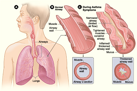
Asthma: This is a chronic disease characterized by inflammation of the airways and constriction of the bronchioles, which can inhibit air from entering the lungs (Fig. 6.7). There is also excessive mucus secretion in the respiratory tract, which contributes to airway occlusion.
Symptoms of asthma include the following:
- Coughing
- Shortness of breath
- Wheezing
- Chest tightness
During a severe asthma attack, the individual may also experience symptoms such as difficultly breathing, cyanosis, confusion, rapid pulse, sweating, and severe anxiety.

Asthma is a very common condition that affects both adults and children. Approximately 8.2% of adults and 9.4% of children in the United States have asthma. It is also the most frequent cause of hospitalization in children.
Atelectasis: This is the collapse of all or part of a lung. It can be caused by pressure on the lung or a blockage of the bronchi or bronchioles (Doyle & McCutcheon, 2020).
Bronchitis: This disease is characterized by inflammation of the lining of the bronchial tubes (Ernstmeyer & Christman, 2020). It often develops from a cold and is a very common pathology. Acute bronchitis, sometimes called a chest cold, usually improves within a week to 10 days without lasting effects, though the cough may linger for weeks. Chronic bronchitis is considered to be a type of chronic obstructive pulmonary disease (COPD) (Ernstmeyer & Christman, 2020).
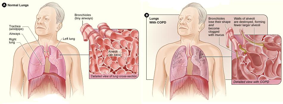
Chronic obstructive pulmonary disease (COPD): This condition is a chronic inflammatory lung disease that causes obstructed airflow out of the lungs (Ernstmeyer & Christman, 2020). It is most often caused by smoking cigarettes, but can also result from long-term exposure to irritating gases or dust. There are two types of COPD: emphysema and chronic bronchitis. Emphysema is a condition in which the alveoli are destroyed and the lungs are hyperinflated (Ernstmeyer & Christman, 2020). Chronic bronchitis, on the other hand, is inflammation of the lining of the bronchial tubes. COPD is treatable but not curable because symptoms often don’t appear until significant lung damage has occurred.
Signs and symptoms of COPD may include the following:
- Shortness of breath, especially during physical activity
- Wheezing
- Chest tightness
- Chronic cough that may produce mucus (sputum) that can be clear, white, yellow, or greenish
- Cyanosis
- Frequent respiratory infections
- Lack of energy
- Unintended weight loss
(Ernstmeyer & Christman, 2020)

Hemoptysis: This condition is characterized by spitting up blood (Chabner, 2018) and can be caused by various respiratory illnesses or trauma.
Hemothorax: This is a collection of blood in the pleural cavity, which is the space between the chest wall and the lung. The most common cause is chest trauma, but hemothorax can also result from abnormal blood clotting or thoracic surgery (Doyle & McCutcheon, 2020).
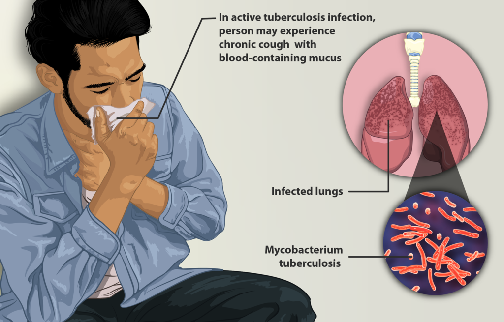
Tuberculosis (TB): The bacteria Mycobacterium tuberculosis causes tuberculosis (TB), a disease that primarily impacts the lungs but can also infect other parts of the body (Ernstmeyer & Christman, 2020). Treatment for TB is challenging and requires patients to take a combination of multiple drugs for an extended time. Some TB strains are multidrug resistant, which makes treatment even more difficult (Ernstmeyer & Christman, 2020).

Exercise
Attribution
Unless otherwise indicated, material on this page has been adapted from the following resource:
Betts, J. G., Young, K. A., Wise, J. A., Johnson, E., Poe, B., Kruse, D. H., Korol, O., Johnson, J. E., Womble, M., & DeSaix, P. (2013). Anatomy and physiology. OpenStax. https://openstax.org/details/books/anatomy-and-physiology licensed under CC BY 4.0
References
Chabner, D. E. (2018). Medical terminology: A short course (8th ed.). Saunders/Elsevier.
CrashCourse. (2015, August 24). Respiratory system, part 1: Crash Course A&P #31 [Video]. YouTube. https://www.youtube.com/watch?v=bHZsvBdUC2I&t=16s
Doyle G. R, & McCutcheon, J. A. (2020). Clinical procedures for safer patient care. BCcampus. https://opentextbc.ca/clinicalskills/ licensed under CC BY 4.0
Ernstmeyer, K., & Christman, E. (Eds.). (2020). Nursing pharmacology. Chippewa Valley Technical College. https://wtcs.pressbooks.pub/pharmacology/ licensed under CC BY 4.0
Image Credits (images are listed in order of appearance)
Human respiratory system-NIH by National Heart, Lung, and Blood Institute, Public domain
Asthma attack-illustration NIH by National Heart, Lung, and Blood Institute, Public domain
Copd 2010Side by National Heart, Lung, and Blood Institute, Public domain
Depiction of a tuberculosis patient by myUpchar, CC BY-SA 4.0
Inflammation of the adenoids
Pertaining to the alveoli
The process of visually examining the bronchial tubes
A chronic condition in which the walls of the bronchi thicken because of inflammation and infection
Inflammation of the bronchioles
Bluish or purplish discolouration caused by oxygen deficiency in the blood
Inflammation of the epiglottis
Inflammation of the larynx
Pertaining to the nose
Inflammation of the nose
Removal of part of the pharynx
The nerve that controls the diaphragm
Inflammation of the pleura
An accumulation of air in the space between the pleura
Inflammation of the lungs
Pertaining to the lungs
Removal of the tonsils
An incision into the trachea

