
3.6 Cardiovascular and Respiratory Systems
Cardiovascular System Overview and Functions
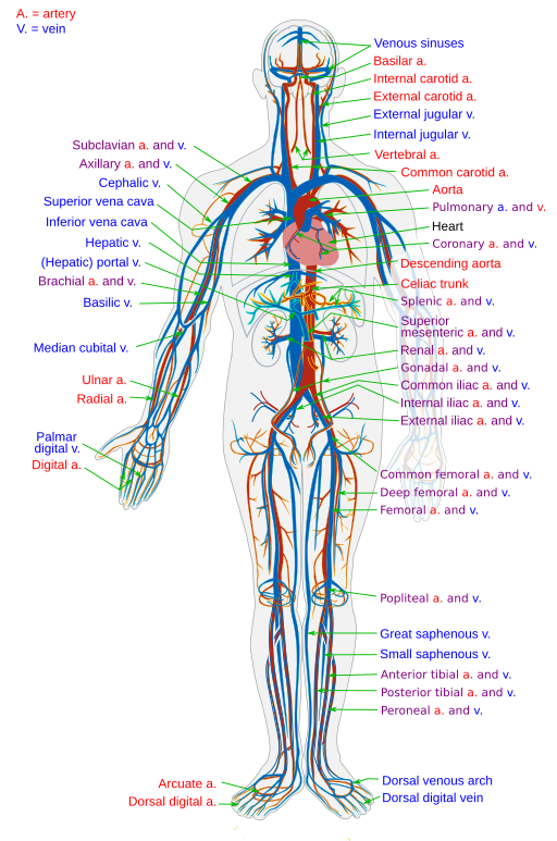
The cardiovascular system (Figure 3.22) uses blood to deliver nutrients and remove wastes from the trillions of cells in the human body. The primary organ in this system, the heart, pumps blood throughout the body via a network of blood vessels. These three components—blood, blood vessels, and the heart—make up this complex system (Study & Erickson, 2022).
Components of the Cardiovascular System
The human heart is located in the thoracic cavity, between the lungs in the space referred to as the mediastinum. The best way to describe the heart is a pump. Its contractions create pressure that sends blood into the major vessels, including the aorta. From there, the blood is distributed to the rest of the body. The vital importance of the heart is obvious. If one assumes an average heart rate of 75 contractions per minute, a human heart would contract approximately 108,000 times in one day, more than 39 million times in one year, and nearly 3 billion times during a 75-year lifespan. Each of the major pumping chambers of the heart ejects approximately 70 millilitres of blood per contraction in a resting adult. This would be equal to 5.25 litres of fluid per minute and approximately 14,000 litres per day. Over one year, that would equal 10 million litres, or 2.6 million gallons, of blood sent through roughly 96,000 kilometres of vessels. To understand how that happens, it is necessary to understand the anatomy and physiology of the heart.
The Heart
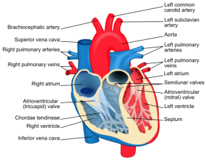
The human heart consists of four chambers—the left side and the right side each have one atrium and one ventricle (Figure 3.23). Each of the upper chambers, the right atrium (plural = atria) and the left atrium, acts as a receiving chamber and contracts to push blood into the lower chambers, the right ventricle and the left ventricle. The ventricles serve as the primary pumping chambers of the heart, propelling blood to the lungs or to the rest of the body.
Blood
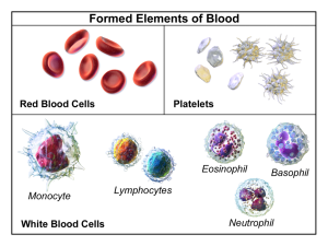
Blood is technically a connective tissue. Like all connective tissues, it is made up of cellular elements and an extracellular matrix. The cellular elements—referred to as the formed elements—include red blood cells (RBCs), white blood cells (WBCs), and cell fragments called platelets (Figure 3.24). The main function of RBCs is to transport oxygen and carbon dioxide to and from the bodies tissues and organs. The primary function of WBCs is to defend the body against infection, and the main function of platelets involves clotting. The extracellular matrix, called plasma, makes blood unique among connective tissues because it is fluid. This fluid, which is mostly water, perpetually suspends the formed elements and enables them to circulate throughout the body within the cardiovascular system.
Blood Vessels
Blood vessels carry blood throughout the body. An artery is a vessel that carries blood away from the heart, where the artery branches into ever-smaller vessels. Eventually, the smallest arteries, called arterioles, further branch into tiny capillaries, where nutrients and wastes are exchanged, and then combine with other vessels that exit capillaries to form venules, small blood vessels that carry blood to a vein, a larger vessel that returns blood to the heart.
The primary function of blood is to deliver oxygen and nutrients to the body’s cells and to remove wastes from those same cells. Blood also has other functions, including defence, distribution of heat, and maintenance of homeostasis.
Common Surgeries Involving the Cardiovascular System
- Angioplasty: Surgical repair of a blood vessel.
- Coronary artery bypass graft (CABG): A procedure done to treat narrowed or blocked arteries by surgically creating a bypass around the blockage. Blood vessels are often taken from the patient’s leg, then used to bypass the blockage in the patient’s chest, thereby creating a new path for blood to travel.
- Carotid endarterectomy: A surgical procedure that removes plaque buildup from the carotid artery to attempt to reduce the chance of a stoke.
- Heart valve repair/replacement: Surgical repair or replacement of a heart valve when it is no longer able to function normally.
- Stent placement: A procedure to place a stent into a newly opened or unblocked blood vessel, which is often done after the treatment of a blockage. The stent stays in place to keep the vessel open and blood flowing.
- Cardiac catheterization: A procedure done to treat or diagnose heart pathologies by inserting a catheter (thin tube) into a blood vessel and up to the patient’s heart.
Respiratory System Overview and Functions
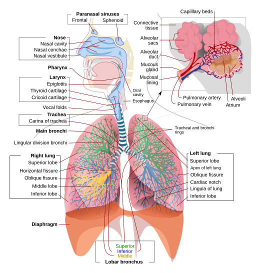
The primary functions of the respiratory system (Figure 3.25) are to provide oxygen to the body’s tissues for cellular respiration, remove the waste product carbon dioxide, and help maintain the acid-base balance (Sturdy & Erickson, 2022). Although cells require oxygen, it is actually the accumulation of carbon dioxide that drives the respiratory system to breathe. This system includes the muscles used to move air in and out of the lungs, the structures involved in the movement of oxygen and carbon dioxide, and the microscopic gas exchange that occurs within the lungs. The majority of the chronic pathologies that affect this system are conditions that impair the gas exchange process and result in laboured breathing and other difficulties (Sturdy & Erickson, 2022).
Components of the Respiratory System
Nose: The nose is the main entrance and exit for the respiratory system.
Pharynx: The pharynx is a tube made up of skeletal muscle and lined with mucous membrane that begins in the nasal cavity and ends at the larynx. The pharynx is divided into three major regions: nasopharynx, oropharynx, and laryngopharynx.
Larynx: This cartilaginous structure is found below the pharynx and connects at the lower end to the trachea. The larynx helps regulate the volume of air that enters and leaves the lungs and is composed of three large cartilage pieces: thyroid cartilage, epiglottis, and cricoid cartilage.
Epiglottis: This very flexible, elastic piece of cartilage covers the opening of the trachea and is attached to the thyroid cartilage. It is an important structure in that it prevents food and liquids from entering the trachea.
Trachea: Also known as the windpipe, the trachea extends from the larynx to the lungs. It branches at the end into the right and left bronchi.
Bronchi: The bronchi lead to tree-like structures in both the right and left lungs that become the smaller bronchioles and finally end in the alveolar sacs and alveoli.
Alveoli: These are the small, almost grape-like structures at the end of the alveolar ducts and are where gas exchange occurs.
Lungs: The lungs are the major organ in the respiratory system and contain the bronchi, bronchioles, and alveoli. The main function of the lungs is to exchange oxygen and carbon dioxide.
Pleura: This serous membrane lines the thoracic cavity and surrounds the lungs. Its purpose is to cushion and protect the lungs.
Diaphragm: This dome-shaped muscle is located at the base of the lungs and divides the thoracic and abdominal cavities. Breathing is dependent on the contraction and relaxation of this muscle.
The lungs exchange respiratory gases across a very large surface area that is highly permeable to gases. This area amazingly totals approximately 70 square metres.
Attribution
Unless otherwise indicated, material on this page has been adapted from the following resource:
Betts, J. G., Young, K. A., Wise, J. A., Johnson, E., Poe, B., Kruse, D. H., Korol, O., Johnson, J. E., Womble, M., & DeSaix, P. (2013). Anatomy and physiology. OpenStax. https://openstax.org/details/books/anatomy-and-physiology, licensed under CC BY 4.0
References
Sturdy, L., & Erickson, S. (2022). The language of medical terminology. Open Education Alberta. https://pressbooks.openeducationalberta.ca/medicalterminology/, licensed under CC BY-NC-SA 4.0
Image Credits
(Images are listed in order of appearance)
Circulatory System by LadyofHats, Public domain
Heart diagram-en by ZooFari, CC BY-SA 3.0
Blausen 0425 Formed Elements by BruceBlaus, CC BY 3.0
Respiratory system complete en by LadyofHats and Jmarchn, Public domain
