
3.5 Nervous System and Sense Organs
Nervous System Overview and Functions
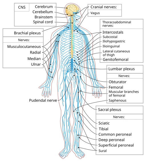
The nervous system (Figure 3.14) is a very complex system and is responsible for controlling much of the body, including both voluntary and involuntary functions (Sturdy & Erickson, 2022). It receives information about the environment around us, and then creates responses to that information. This system is also responsible for taking sensory input and integrating it with other sensations, memories, emotional states, and learning. The nervous system can be divided into two main components: the central nervous system and the peripheral nervous system. From there, it is further subdivided by functions and components (Sturdy & Erickson, 2022).
Components of the Nervous System
The two main components of the nervous system are the central nervous system (CNS) and the peripheral nervous system (PNS).
The CNS is made up of the brain and the spinal cord. The regulation of homeostasis and conscious experiences are controlled in the brain (Sturdy & Erickson, 2022).
Key Concepts
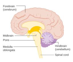
The brain is is made up of its major regions, which include the cerebrum, diencephalon, brain stem, and cerebellum (Figure 3.15).
The cerebrum is the iconic grey mantle of the human brain, which appears to make up most of the mass of the brain. Many of the higher neurological functions such as memory, emotion, and consciousness are the result of cerebral function.
The diencephalon is the one region of the brain that retains its name from embryologic development. It is the connection between the cerebrum and the rest of the nervous system, with one exception. The rest of the brain, the spinal cord, and the PNS all send information to the cerebrum through the diencephalon, and output from the cerebrum passes through the diencephalon. The single exception is the system associated with olfaction, or the sense of smell, which connects directly with the cerebrum.
The midbrain and hindbrain (composed of the pons and the medulla) are collectively referred to as the brain stem. The structure emerges from the ventral surface of the forebrain as a tapering cone that connects the brain to the spinal cord. Attached to the brain stem, but considered a separate region of the adult brain, is the cerebellum. The midbrain coordinates sensory representations of the visual, auditory, and somatosensory perceptual spaces. The pons is the main connection with the cerebellum. The pons and the medulla regulate several crucial functions, including the cardiovascular and respiratory systems and rates.
The cerebellum is largely responsible for comparing information from the cerebrum with sensory feedback from the periphery through the spinal cord. It accounts for approximately 10% of the mass of the brain.
The spinal cord is another major organ of the system. Whereas the brain develops out of expansions of the neural tube into primary and then secondary vesicles, the spinal cord maintains the tube structure and is only specialized into certain regions. On the whole, the posterior regions are responsible for sensory functions, and the anterior regions are associated with motor functions.
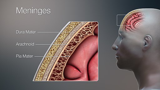
The outer surface of the CNS is covered by a series of three membranes composed of connective tissue called the meninges, which protect the brain (Figure 3.16):
- The dura mater is a thick, fibrous layer and a strong protective sheath over the entire brain and spinal cord. It is anchored to the inner surface of the cranium and vertebral cavity.
- The arachnoid mater is a membrane of thin, fibrous tissue that forms a loose sac around the CNS. Beneath the arachnoid is a thin, filamentous mesh called the arachnoid trabeculae, which looks like a spiderweb, giving this layer its name.
- Directly adjacent to the surface of the CNS is the pia mater, a thin, fibrous membrane that follows the convolutions of the gyri and sulci in the cerebral cortex and fits into other grooves and indentations.
The peripheral nervous system (PNS) connects the central nervous system with the rest of the body. The nerves, axons, and ganglia that make up the PNS are found throughout the body, including in other organs and even in other systems, such as the digestive system, as well as the eyes, ears, nose, and various other locations. Messages travel back and forth from the CNS to the muscles, organs, and senses in the peripheral areas of the body via the PNS. The sensory neurons that carry messages and various forms of sensory information towards the CNS are considered afferent fibres. When the CNS uses motor neurons to carry instructions from the CNS to the muscles, they are called efferent fibres. Messages continually go back and forth along neurons between the CNS and the periphery. The PNS has three subdivisions as well—the enteric nervous system, somatic nervous system, and autonomic nervous system:
- Somatic nervous system: This part of the PNS is responsible for conscious perception of the environment and for voluntary responses to that perception through the use of skeletal muscles (Sturdy & Erickson, 2022).
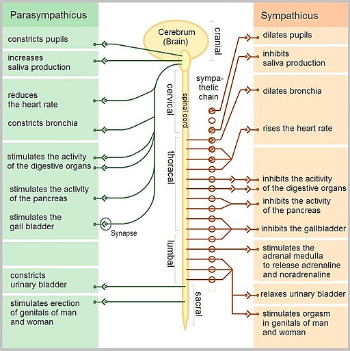
- Autonomic nervous system: This part of the PNS handles involuntary responses that the brain controls without the need for conscious thought. It consists of the sympathetic and parasympathetic nervous systems and uses a balance of the two to regulate the body’s involuntary functions, including heart rate, respiratory rate, digestion, and sweating:
- Sympathetic nervous system: Associated with the fight-or-flight response
- Parasympathetic nervous system: Focuses on what could be called “rest and digest”
(Sturdy & Erickson, 2022)
- Enteric nervous system (ENS): This part of the PNS is responsible for controlling the smooth muscle and glandular tissue in your digestive system. It is a large part of the PNS and is not dependent on the CNS. This part of the PNS is often considered part of the autonomic nervous system, though some resources list it as separate because of slight variations between the two.
Figure 3.17 shows how the sympathetic and parasympathetic nervous systems work in the body. Both affect the same areas of the body but in a different manner.
Sense Organs
A major role of sensory receptors is to help us learn about the environment around us or about the state of our internal environment. Stimuli from varying sources, and of different types, are received and changed into the electrochemical signals of the nervous system. This occurs when a stimulus changes the cell membrane potential of a sensory neuron. The stimulus causes the sensory cell to produce an action potential that is relayed into the central nervous system (CNS), where it is integrated with other sensory information, or sometimes higher cognitive functions, to become a conscious perception of that stimulus. Below we will discuss four of the main sense organs in the human body. Although there are more, these are the most common and are applicable to your future role.
Gustation (Taste)
Gustation, or taste, is the special sense associated with the tongue. The surface of the tongue, along with the rest of the oral cavity, is lined by a stratified squamous epithelium. Raised bumps called papillae (singular = papilla) contain the structures for gustatory transduction. There are four types of papillae, based on their appearance: circumvallate, foliate, filiform, and fungiform (Figure 3.18). Within the structure of the papillae are taste buds that contain specialized gustatory receptor cells for the transduction of taste stimuli. These receptor cells are sensitive to the chemicals contained within foods that are ingested, and they release neurotransmitters based on the amount of the chemicals in the food. Neurotransmitters from the gustatory cells can activate sensory neurons in the facial, glossopharyngeal, and vagus cranial nerves.
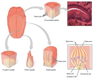
Only a few recognized sub-modalities exist within the sense of gustation. Until recently, only four tastes were recognized: sweet, salty, sour, and bitter. Research at the turn of the 20th century led to recognition of a fifth taste, umami, during the mid-1980s. Umami is a Japanese word that means “delicious taste” and is often translated to mean “savoury.” Very recent research has suggested that there may also be a sixth taste for fats, or lipids.
Olfaction (Smell)
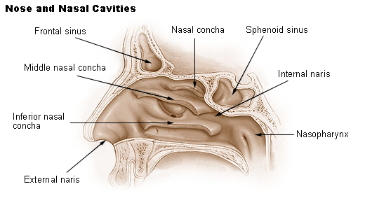
Like taste, the sense of smell, or olfaction, is also responsive to chemical stimuli. Figure 3.19 is an illustration of the main components of the nose and nasal cavities. The olfactory receptor neurons are located in a small region in the superior nasal cavity. This region is referred to as the olfactory epithelium and contains bipolar sensory neurons. Each olfactory sensory neuron has dendrites that extend from the apical surface of the epithelium into the mucus lining the cavity. As airborne molecules are inhaled through the nose, they pass over the olfactory epithelial region and dissolve into the mucus. These odorant molecules bind to proteins that keep them dissolved in the mucus and help transport them to the olfactory dendrites. The odorant–protein complex binds to a receptor protein within the cell membrane of an olfactory dendrite. These receptors are G protein–coupled and will produce a graded membrane potential in the olfactory neurons.
Audition (Hearing)
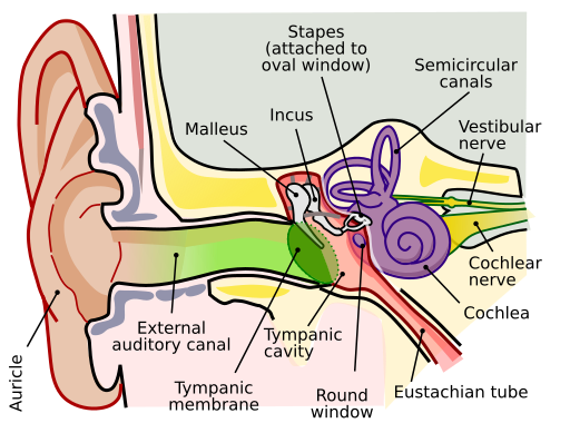
Hearing, or audition, is the transduction of sound waves into a neural signal that is made possible by the structures of the ear (Figure 3.20) The large, fleshy structure on the lateral aspect of the head is known as the auricle. The C-shaped curves of the auricle direct sound waves toward the auditory canal. At the end of the auditory canal is the tympanic membrane, or eardrum, which vibrates after it is struck by sound waves. The auricle, ear canal, and tympanic membrane are often referred to as the external ear.
The middle ear consists of a space spanned by three small bones called the ossicles. The three ossicles are the malleus, incus, and stapes, which are Latin names that roughly translate to hammer, anvil, and stirrup. The malleus is attached to the tympanic membrane and articulates with the incus. The incus, in turn, articulates with the stapes. The stapes is attached to the inner ear, where the sound waves are transduced into a neural signal. The middle ear is connected to the pharynx through the Eustachian tube, which helps equalize pressure across the tympanic membrane.
Vision
Vision is the special sense of sight that is based on the transduction of light stimuli received through the eyes. The eyes are located within either orbit in the skull. The bony orbits surround the eyeballs, protecting them and anchoring the soft tissues of the eye (Figure 3.21). The eyelids, with lashes at their leading edges, help to protect the eye from abrasions by blocking particles that may land on the surface of the eye. The inner surface of each lid is a thin membrane known as the palpebral conjunctiva. The conjunctiva extends over the white areas of the eye (the sclera), connecting the eyelids to the eyeball. Tears are produced by the lacrimal gland, located just inside the orbit, superior and lateral to the eyeball. Tears produced by this gland flow through the lacrimal duct to the medial corner of the eye, where the tears flow over the conjunctiva, washing away foreign particles.
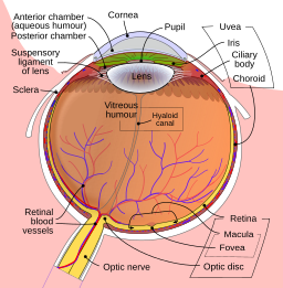
The eyeball itself is a hollow sphere composed of three layers of tissue:
- The outermost layer is the fibrous tunic, which includes the white sclera and clear cornea. The sclera accounts for five-sixths of the surface of the eye, most of which is not visible, though humans are unique compared with many other species in having so much of the “white of the eye” visible. The transparent cornea covers the anterior part of the eye. It allows light to enter and, along with the lens, focuses light onto the retina.
- The middle layer of the eye is the vascular tunic, which is mostly composed of the choroid, ciliary body, and iris. The choroid is a layer of highly vascularized connective tissue that supplies blood to the eyeball. The choroid is posterior to the ciliary body, a muscular structure attached to the lens by suspensory ligaments, or zonule fibres. These two structures bend the lens, allowing it to focus light on the back of the eye. Overlaying the ciliary body, and visible in the anterior eye, is the iris—the coloured part of the eye. The iris is a smooth muscle that opens or closes the pupil, which is the hole at the centre of the eye that allows light to enter. The iris constricts the pupil in response to bright light and dilates the pupil in response to dim light.
- The innermost layer of the eye is the neural tunic, or retina, which contains the nervous tissue responsible for photoreception.
The eye is also divided into two cavities: the anterior cavity and the posterior cavity. The anterior cavity is the space between the cornea and lens, including the iris and ciliary body. It is filled with a watery fluid called the aqueous humour. The posterior cavity is the space behind the lens that extends to the posterior side of the interior eyeball, where the retina is located. The posterior cavity is filled with a more viscous fluid called the vitreous humour.
The retina is composed of several layers and contains specialized cells for the initial processing of visual stimuli. The photoreceptors (rods and cones) change their membrane potential when stimulated by light energy. The change in membrane potential alters the amount of neurotransmitter that the photoreceptor cells release onto bipolar cells in the outer synaptic layer. It is the bipolar cell in the retina that connects a photoreceptor to a retinal ganglion cell (RGC) in the inner synaptic layer. There, amacrine cells additionally contribute to retinal processing before an action potential is produced by the RGC. The axons of RGCs, which lie at the innermost layer of the retina, collect at the optic disc and leave the eye as the optic nerve. Because these axons pass through the retina, there are no photoreceptors at the very back of the eye, where the optic nerve begins. This creates a “blind spot” in the retina, and a corresponding blind spot in our visual field.
Common Surgeries Involving the Nervous System
- Craniotomy: This is an incision, or opening, into the skull. This procedure can be done as part of brain surgery or as a means to relieve pressure in the skull (Sturdy & Erickson, 2022).
- Lumbar puncture (LP): Also referred to as a spinal tap, this procedure is done to take a sample of cerebrospinal fluid to send to the lab for analysis (Sturdy & Erickson, 2022).
- Neurosurgery: This general term relates to any surgical procedure involving the nervous system.
- Cataract surgery and glaucoma surgery: These are common procedures performed on the eye to treat either cataracts or glaucoma.
- Tympanoplasty: Surgical repair of the tympanic membrane, or eardrum.
Attribution
Unless otherwise indicated, material on this page has been adapted from the following resource:
Betts, J. G., Young, K. A., Wise, J. A., Johnson, E., Poe, B., Kruse, D. H., Korol, O., Johnson, J. E., Womble, M., & DeSaix, P. (2013). Anatomy and physiology. OpenStax. https://openstax.org/details/books/anatomy-and-physiology, licensed under CC BY 4.0
References
Sturdy, L., & Erickson, S. (2022). The language of medical terminology. Open Education Alberta. https://pressbooks.openeducationalberta.ca/medicalterminology/, licensed under CC BY-NC-SA 4.0
Image Credits
(Images are listed in order of appearance)
Nervous system diagram-en by Medium69 and Jmarchn, CC BY-SA 4.0
Diagram showing the brainstem which includes the medulla oblongata, the pons and the midbrain (2) CRUK 294 by Cancer Research UK, CC BY-SA 4.0
3D Medical Illustration Meninges Details by Scientific Animations, CC BY-SA 4.0
The Autonomic Nervous System by Geo-Science-International, CC0 1.0 Public domain
1402 The Tongue by OpenStax, CC BY 4.0
Nose and nasal cavities by National Cancer Institute SEER Training, Public domain
Anatomy of the Human Ear. From Chittka, L., & Brockmann, A. (2005). Perception space – The final frontier. PLoS Biology, 3(4), e137. https://doi.org/10.1371/journal.pbio.0030137
Schematic diagram of the human eye en by Rhcastilhos and Jmarchn, CC BY-SA 3.0

_CRUK_294.svg)