
3.3 Skeletal and Muscular Systems
Skeletal System Overview and Functions
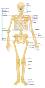
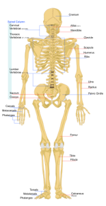
The skeletal system consists of all the bones, joints, tendons, and cartilage found in the human body (Figures 3.6 and 3.7). Bone, or osseous tissue, is a hard, dense connective tissue that forms most of the adult skeleton, the support structure of the body. In the areas of the skeleton where bones move (for example, the rib cage and joints), cartilage, a semi-rigid form of connective tissue, provides flexibility and smooth surfaces for movement. The skeletal system is the body system composed of bones and cartilage and performs the following critical functions for the human body:
- Supports the body
- Facilitates movement
- Protects internal organs
- Produces blood cells
- Stores and releases minerals and fat
Components of the Skeletal System
The skeleton is subdivided into two major components:
- The axial skeleton forms the vertical, central axis of the body and includes all the bones of the head, neck, chest, and back. It protects the brain, spinal cord, heart, and lungs. It also serves as the attachment site for muscles that move the head, neck, and back, and for muscles that act across the shoulder and hip joints to move their corresponding limbs. There are 80 bones in the axial skeleton.
- The appendicular skeleton includes all the bones of the upper and lower limbs, plus the bones that attach each limb to the axial skeleton. There are 126 bones in the appendicular skeleton.
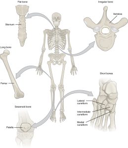
In total, 206 bones compose the adult skeleton. The shapes and functions of the bones are related such that each categorical shape of bone has a distinct function. Bones are classified as either long, short, flat, irregular, or sesamoid. Table 3.1 and Figure 3.8 provide details about each bone type.
| Bone Classification | Features | Function(s) | Examples |
|---|---|---|---|
| Long | Cylinder-like shape; longer than it is wide | Leverage | Femur, tibia, fibula, metatarsals, humerus, ulna, radius, metacarpals, phalanges |
| Short | Cube-like shape; approximately equal in length, width, and thickness | Provide stability and support while allowing some motion | Carpals, tarsals |
| Flat | Thin and curved | Points of attachment for muscles; protect internal organs | Sternum, ribs, scapulae, cranial bones |
| Irregular | Complex shape | Protect internal organs | Vertebrae, facial bones |
| Sesamoid | Small and round; embedded in tendons | Protect tendons from compressive forces | Patellae |
Bones
Bone is the hardest connective tissue. It provides protection to internal organs and supports the body. The rigid extracellular matrix of bones contains mostly collagen fibres embedded in a mineralized ground substance containing hydroxyapatite, a form of calcium phosphate. Both components of the matrix, organic and inorganic, contribute to the unusual properties of bone. Without collagen, bones would be brittle and shatter easily. Without mineral crystals, bones would flex and provide little support.
There are also both cancellous and compact types of bones. Under a microscope, cancellous bone looks like a sponge and has empty spaces between trabeculae, or arches of bone proper. It is lighter than compact bone and is found in the interior of some bones and at the ends of long bones. Compact bone is solid and has greater structural strength than cancellous bone.
Joints
Joints, also known as articulation, are any place where adjacent bones or bone and cartilage come together to form a connection. Many joints allow for movement between bones. At these joints, the articulating surfaces of the adjacent bones can move smoothly against each other; however, the bones of other joints may be joined by connective tissue or cartilage. These joints are designed for stability and allow little or no movement. Importantly, joint stability and movement are related to each other—stable joints have little or no mobility between the adjacent bones, whereas joints that provide the most movement between bones are the least stable. Understanding the relationship between joint structure and function helps explain why particular types of joints are found in certain areas of the body.
The structural classification of joints is based on whether the articulating surfaces of the adjacent bones are directly connected by fibrous connective tissue or cartilage, or whether the articulating surfaces contact each other within a fluid-filled joint cavity. These differences serve to divide the joints of the body into three structural classifications:
- At a fibrous joint, adjacent bones are united by fibrous connective tissue.
- At a cartilaginous joint, the bones are joined by hyaline cartilage or fibrocartilage.
- At a synovial joint, the articulating surfaces of the bones are not directly connected, but instead come into contact with each other within a joint cavity that is filled with a lubricating fluid. Synovial joints allow for free movement between the bones and are the most common joints in the body.
Synovial joints are subdivided based on the shapes of the articulating surfaces of the bones that form each joint. The six types of synovial joints are pivot, hinge, condyloid, saddle, plane, and ball-and-socket joints:
- At a pivot joint, a rounded portion of bone is enclosed within a ring formed partially by the articulation with another bone and partially by a ligament. The bone rotates within this ring. The pivot joint in your neck (between the atlas and the axis) allows you to rotate your head.
- In a hinge joint, the convex end of one bone articulates with the concave end of the adjoining bone. This type of joint allows only for bending and straightening motions along a single axis, and hinge joints are functionally classified as uniaxial joints. The knees and elbows are hinge joints.
- At a condyloid joint (ellipsoid joint), the shallow depression at the end of one bone articulates with a rounded structure from an adjacent bone or bones. The knuckle (metacarpophalangeal) joints of the hand between the distal end of a metacarpal bone and the proximal phalanx bone are condyloid joints.
- At a saddle joint, both of the articulating surfaces of the bones have a saddle shape, which is concave in one direction and convex in the other. This allows the two bones to fit together like a rider sitting on a saddle. Saddle joints are functionally classified as biaxial joints. The joint where the thumb joins the hand is a saddle joint.
- At a plane joint (gliding joint), the articulating surfaces of the bones are flat or slightly curved and of approximately the same size, which allows the bones to slide against each other. The motion at this type of joint is usually small and tightly constrained by surrounding ligaments. Based only on their shape, plane joints can allow multiple movements, including rotation. Plane joints can be found in the wrists and ankles, as well as in other parts of the body.
- The joint with the greatest range of motion is the ball-and-socket joint. At these joints, the rounded head of one bone (the ball) fits into the concave articulation (the socket) of the adjacent bone. The hip joint and the shoulder joint are the only ball-and-socket joints in the body.
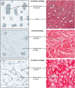
Cartilage is elastic connective tissue found at the ends of bones as well as in other locations such as the tip of the nose. The distinctive appearance of cartilage is caused by polysaccharides called chondroitin sulfates, which bind with ground substance proteins to form proteoglycans. The three main types of cartilage tissue are hyaline cartilage, fibrocartilage, and elastic cartilage (Figure 3.9):
- Hyaline cartilage, the most common type of cartilage in the body, consists of short, dispersed collagen fibres and contains large amounts of proteoglycans. The surface of hyaline cartilage is smooth, and it is found in the rib cage and nose and covers bone ends where they meet to form moveable joints. It makes up a template of the embryonic skeleton before bone formation. A plate of hyaline cartilage at the ends of bones allows for continued growth until adulthood.
- Fibrocartilage is tough because it has thick bundles of collagen fibres dispersed throughout its matrix. Menisci in the knee joints and intervertebral discs are examples of fibrocartilage.
- Elastic cartilage contains elastic fibres as well as collagen and proteoglycans. This tissue gives rigid support as well as elasticity.
Ligaments and tendons are made of dense regular connective tissue, but in ligaments, not all the fibres are parallel. Dense regular elastic tissue contains elastin fibres in addition to collagen fibres, which allows the ligament to return to its original length after stretching. The ligaments in the vocal folds and between the vertebrae in the vertebral column are elastic.
Common Surgeries Involving the Skeletal System
- Open reduction internal fixation (ORIF) involves repairing a fracture by exposing the site of the injury, realigning the bone, and then stabilizing it with wires, plates, and screws (Sturdy & Erickson, 2022).
- Joint replacements are commonly performed, and the site of the surgery varies. Hip arthroplasty and knee arthroplasty are the most common surgeries and involve replacement of the articular surfaces of those joints.
- Investigative surgical procedures are also commonly completed; the medical term for this procedure is arthroscopy (Sturdy & Erickson, 2022). It involves using instruments to view what is inside a joint.
- Resections on the skeletal system include surgeries such as vertebrectomy and meniscectomy (Sturdy & Erickson, 2022).
- Osteotomy involves cutting and realigning bones and joints. This procedure can also be done to correct deformities or misalignment (Sturdy & Erickson, 2022).
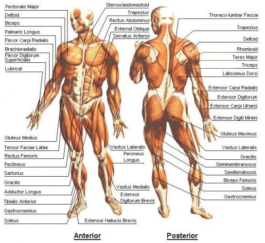
Muscular System Overview and Functions
The muscular system consists of all the muscles in the human body (Figure 3.10). The purpose of this system is to support movement of the body and to protect and assist with the functions of other body systems; for example, cardiac muscles help the heart beat (Sturdy & Erickson, 2022).
Components of the Muscular System
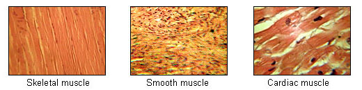
Muscle is one of the four primary tissue types of the body, and the body has three kinds of muscle tissue: skeletal muscle, smooth muscle, and cardiac muscle (Figure 3.11) (Sturdy & Erickson, 2022). All three muscle tissues have some properties in common; for example, they all exhibit a quality called “excitability” because their plasma membranes can change their electrical state (from polarized to depolarized) and send an electrical wave called an “action potential” along the entire length of the membrane. Although the nervous system can influence the excitability of cardiac and smooth muscle to some degree, skeletal muscle completely depends on signalling from the nervous system to work properly. On the other hand, both cardiac muscle and smooth muscle can respond to other stimuli, such as hormones and local stimuli.
The best-known feature of skeletal muscle is its ability to contract and cause movement. Skeletal muscles act not only to produce movement, but also to stop movement, such as resisting gravity to maintain posture. Small, constant adjustments of the skeletal muscles are needed to hold a body upright or balanced in any position. Skeletal muscles are located throughout the body at the openings of internal tracts to control the movement of various substances. These muscles allow functions such as swallowing, urination, and defecation to be under voluntary control. Skeletal muscles also protect internal organs, particularly abdominal and pelvic organs, by acting as an external barrier or shield to external trauma and by supporting the weight of the organs.
Smooth muscle (sometimes called involuntary muscle) is present in the walls of hollow organs such as the urinary bladder, uterus, stomach, and intestines, and in the walls of passageways, such as the arteries and veins of the circulatory system and the tracts of the respiratory, urinary, and reproductive systems. Smooth muscle is also present in the eyes, where it functions to change the size of the iris and alter the shape of the lens, and in the skin, where it causes hair to stand erect in response to cold temperatures or fear.
Cardiac muscle is only found in the heart. Highly coordinated contractions of cardiac muscle pump blood into the vessels of the circulatory system.
Common Surgeries Involving the Muscular System
- The majority of surgeries that involve the muscular system are performed because of injuries or conditions related to the skeletal system as well. Surgery can also be done to repair muscles, or in some cases, a procedure called a fasciotomy might be performed.
Attribution
Unless otherwise indicated, material on this page has been adapted from the following resource:
Betts, J. G., Young, K. A., Wise, J. A., Johnson, E., Poe, B., Kruse, D. H., Korol, O., Johnson, J. E., Womble, M., & DeSaix, P. (2013). Anatomy and physiology. OpenStax. https://openstax.org/details/books/anatomy-and-physiology, licensed under CC BY 4.0
References
Sturdy, L., & Erickson, S. (2022). The language of medical terminology. Open Education Alberta. https://pressbooks.openeducationalberta.ca/medicalterminology/, licensed under CC BY-NC-SA 4.0
Image Credits
(Images are listed in order of appearance)
Human skeleton front en by LadyofHats, Public domain
Human skeleton back en by LadyofHats, Public domain
Figure 6.6 Classification of Bones. From Betts, J. G., Young, K. A., Wise, J. A., Johnson, E., Poe, B., Kruse, D. H., Korol, O., Johnson, J. E., Womble, M., & DeSaix, P. (2013). 6.2 Bone classification. In Anatomy and physiology. OpenStax. https://openstax.org/details/books/anatomy-and-physiology, licensed under CC BY 4.0
Figure 4.16 Types of Cartilage. From Betts, J. G., Young, K. A., Wise, J. A., Johnson, E., Poe, B., Kruse, D. H., Korol, O., Johnson, J. E., Womble, M., & DeSaix, P. (2013). 4.3 Connective tissue supports and protects. In Anatomy and physiology. OpenStax. https://openstax.org/details/books/anatomy-and-physiology, licensed under CC BY 4.0
Human Body-Muscular by Unknown, CC BY-SA 4.0
Illu muscle tissues by National Cancer Institute SEER Training, Public domain
Removal of a vertebra
Removal of the cartilage pad within the knee joint
A procedure that involves cutting through the connective tissue that wraps around muscles in order to treat a condition called compartment syndrome
