
3.4 Integumentary and Lymphatic Systems
Integumentary System Overview and Functions
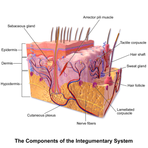
The integumentary system (Figure 3.12) is one of the largest body systems and makes up approximately 16% of total body weight (Sturdy & Erickson, 2022). The main component of this system is the skin, which is responsible for much more than simply contributing to your outward appearance. The skin protects the inner organs, controls thermoregulation, functions as a sensory organ, and is necessary for vitamin D synthesis. It is made up of multiple layers of cells and tissues that are held to underlying structures by connective tissues (Sturdy & Erickson, 2022). The deepest layer of skin has many blood vessels and also has sensory, autonomic, and sympathetic nerve fibres that ensure communication to and from the central nervous system (CNS). Accessory organs within this system include hair, nails, and various glands.
Components of the Integumentary System
Epidermis: This is the outermost skin layer and is composed of keratinized, stratified squamous epithelium. The epidermis is made of four or five layers of epithelial cells, depending on its location in the body. It is avascular and does not contain any blood vessels.
Dermis: This inner skin layer is the main component of the integumentary system and contains blood and lymph vessels, nerves, and other structures, such as hair follicles and sweat glands. The dermis is made of two layers of connective tissue that compose an interconnected mesh of elastin and collagenous fibres, which are produced by fibroblasts.
Hypodermis: This layer is also known as the subcutaneous layer and lies below the dermis. It connects the skin to the fibrous tissues of the bones and muscles.
Hair: This is a keratinous filament that grows out of the epidermis. It is primarily made up of dead, keratinized cells.
Nail bed: This epidermal structure is found at the tips of our fingers and toes. The nail body is formed on the nail bed and protects the tips of our fingers and toes and helps us pick up small objects.
Sudoriferous glands: Also known as sweat glands, these glands produce sweat to cool the body when it becomes warm. There are two types of sweat glands, and each secretes slightly different products.
Sebaceous glands: These glands are a type of oil gland and are found all over the body. They help to lubricate and waterproof the skin and hair. Many of these glands are found near hair follicles. The sebaceous glands generate and excrete sebum, which is a mixture of lipids, onto the skin surface, lubricating the dry and dead layer of keratinized cells. The secretion of sebum is stimulated by hormones, many of which do not become active until puberty.
Common Surgeries Involving the Integumentary System
- Biopsies are a common procedure that might involve this body system. A sample of skin or other matter is taken and then sent to the lab for analysis (Sturdy & Erickson, 2022).
- A skin graft, which can be partial or full thickness, involves taking skin from a donor site and placing it somewhere else on the body.
Lymphatic System Overview and Functions
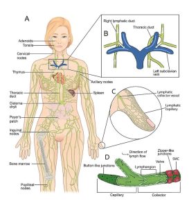
The lymphatic system (Figure 3.13) is one of the lesser-known systems of the human body, but it has an extremely important function. It consists of a network of lymphatic vessels, lymph nodes, and ducts (Sturdy & Erickson, 2022). One of the major functions of the lymphatic system is to drain body fluids and return them to the bloodstream. Blood pressure can cause leakage of fluid from the capillaries, resulting in the accumulation of fluid in the spaces between individual cells in the tissues. Each day, about 20 litres of plasma flow through the body’s arteries, arterioles, and capillaries, but only about 17 litres are returned to circulation by way of the veins (Sturdy & Erickson, 2022). The rest is released into the interstitial spaces of the tissues because of capillary filtration. The lymphatic system collects this excess fluid, now called lymph, from tissues in the body and returns it to the bloodstream (Sturdy & Erickson, 2022).
The lymphatic system is also associated with the immune system to the point that the systems are virtually indistinguishable (Sturdy & Erickson, 2022). The immune system is the complex collection of cells and organs that destroy and/or neutralize pathogens that could cause disease or death. Cells in the immune system use lymphatic vessels to make their way from the interstitial spaces back into the circulatory system, and they also use lymph nodes as staging areas for the development of an immune response. For example, lymph nodes swell during an infection, and lymphocytes are transported via the lymphatic vessels.
Components of the Lymphatic System
Lymph: Lymph is the fluid contained within the lymphatic system.
Lymphatic vessels: These vessels begin as capillaries, which feed into larger and larger lymphatic vessels, and eventually empty into the bloodstream through a series of ducts.
Lymphatic capillaries: These are the smallest of the lymphatic vessels and the start of the lymph flow. Interstitial fluid enters the lymphatic system via the capillaries to become lymph fluid. Lymphatic capillaries are located in almost every tissue in the body and are interwoven among the arterioles and venules of the circulatory system.
Lymphatic trunks: These are large lymphatic vessels that collect lymph from the smaller lymphatic vessels, such as the capillaries, and empty it into the blood via lymphatic ducts.
Lymphocytes: These are the primary cells of adaptive immune responses. The two basic types of lymphocytes are B cells and T cells, which are distinguished from one another by their surface protein markers, as well as by the molecules they secrete. Both types of cells initially develop in the bone marrow, but then the T cells move to the thymus to mature and the B cells remain in the bone marrow.
The primary lymphoid organs are where lymphocytes mature, proliferate, and are selected, which enables them to attack pathogens without harming the cells of the body. The primary lymphoid organs are the bone marrow and the thymus gland:
- Bone marrow: This primary lymphoid organ is where B cells undergo nearly all of their development. It is also where immature T cells develop until they move to the thymus gland.
- Thymus gland: This primary lymphoid organ is found in the space between the sternum and the aorta. It is involved in the development and maturing of T cells and is most active during infancy and childhood.
Lymphocytes develop and mature in the primary lymphoid organs, but they mount immune responses from the secondary lymphoid organs. A naïve lymphocyte is one that has left the primary organ and entered a secondary lymphoid organ. Naïve lymphocytes are fully functional immunologically but have yet to encounter an antigen to respond to. In addition to circulating in the blood and lymph, lymphocytes concentrate in secondary lymphoid organs. Secondary lymphoid organs include the spleen, lymph nodes, and lymphoid nodes:
- Spleen: The spleen is considered to be a major secondary lymphoid organ. It is about 12 centimetres long and is a fragile organ without a strong capsule. The spleen is sometimes called the “filter of the blood” because of its extensive vascularization and the fact that it removes microbes and other materials from the blood.
- Lymph nodes: These bean-shaped organs can be found throughout the body. Humans have 500 to 600 lymph nodes. Their function is to remove debris and pathogens from the lymph. Bacteria that infect interstitial fluid are taken up by the lymphatic capillaries and transported to a regional lymph node.
- Lymphoid nodules: These nodules are non-encapsulated patches of lymphoid tissue and are found throughout the body. They are simpler than the lymph nodes and spleen in that they consist simply of a dense cluster of lymphocytes without a surrounding fibrous capsule. These nodules are located in the respiratory and digestive tracts, which are areas routinely exposed to environmental pathogens.
- Tonsils: These lymphoid nodules are located on the inner surface of the pharynx. They are important in fighting oral pathogens and developing immunity.
Common Surgeries Involving the Lymphatic System
Attribution
Unless otherwise indicated, material on this page has been adapted from the following resource:
Betts, J. G., Young, K. A., Wise, J. A., Johnson, E., Poe, B., Kruse, D. H., Korol, O., Johnson, J. E., Womble, M., & DeSaix, P. (2013). Anatomy and physiology. OpenStax. https://openstax.org/details/books/anatomy-and-physiology, licensed under CC BY 4.0
References
Sturdy, L., & Erickson, S. (2022). The language of medical terminology. Open Education Alberta. https://pressbooks.openeducationalberta.ca/medicalterminology/, licensed under CC BY-NC-SA 4.0
Image Credits
(Images are listed in order of appearance)
Blausen 0810 SkinAnatomy 01 by BruceBlaus, CC BY 3.0
Anatomy of the lymphatic system by SGUL lymres, CC BY-SA 4.0
