
Glossary
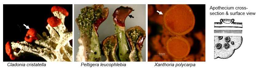
Apothecium (plural: apothecia)
Disc or cup-shaped structures formed by the fungal symbiont, where sexual spores are created. The spore-containing surface is usually a different colour than the thallus of the lichen. Line drawing reproduced courtesy of BC Ministry of Forests and T. Goward, from Goward et al. 1994, fig. 9a.
Bipartite (adjective)
A lichen that is composed of two dominant partners, at least one fungus (=mycobiont) and an alga (=photobiont). Most lichens, both chlorolichens and cyanolichens, are bipartitate. Compare with tripartite.
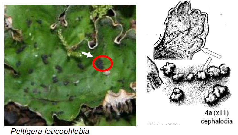
Cephalodium (plural: cephalodia)
Colony of cyanobacteria on a lichen with a primarily-green algal photobiont. When occurring on the surface, they appear as small, brain-shaped or wart-like structures. Vary in shape and degree of attachment. Line drawing reproduced courtesy of BC Ministry of Forests and T. Goward, from Goward et al. 1994, Key to Peltigera and Similar Lichens, p. 97.
Chlorolichen (plural: chlorolichens)
Lichens where the dominant photosynthetic partner is a green alga like Trebouxia.
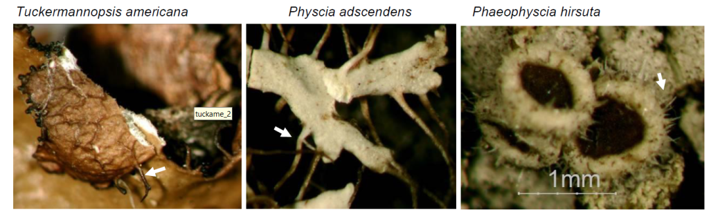
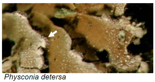
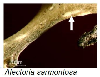
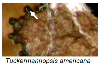
Cilium (plural: cilia)
Hair-like structures, arising from lobe margins and tips, sometimes from apothecial margins.
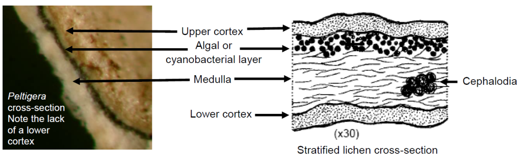
Cortex (plural: cortices)
Outer protective layer of a lichen thallus, composed of fungal cells embedded in a matrix. Many foliose lichens have differentiated upper and lower cortices, while some lichens have only an upper cortex (e.g., Peltigera), and fruticose lichens typically have one homogenous cortex. Line drawing reproduced courtesy of BC Ministry of Forests and T. Goward, from Goward et al. 1994, fig. 4.
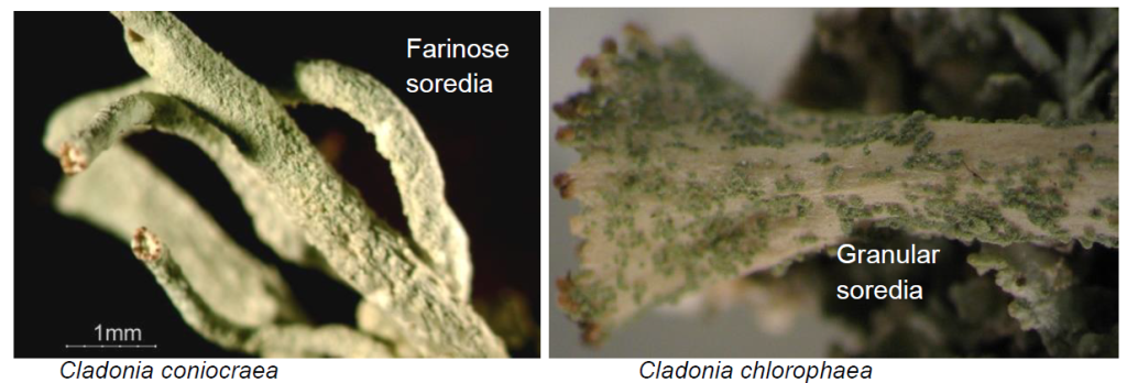
Farinose
Fine and powdery or chalky (flour-like), as compared to coarse, granular (salt-like). Describes soredia.
Foliose (adjective, growth form)
Mostly growing in 2-dimensions, typically flat and leaf-life, usually has a distinct and separate upper and lower cortex (but some genera lack a lower cortex); typically with multiple attachments points but can be detached from the substrate intact and may even sparsely attached or unattached. See entry for growth forms.
Fruticose (adjective, growth form)
Clearly 3-dimensional, one cortex surrounds thallus (but may be ecorticate), often branched, may resemble shrubs, hair or stick-pins; easily detached from substrate with very limited attachment points. See entry for growth forms.
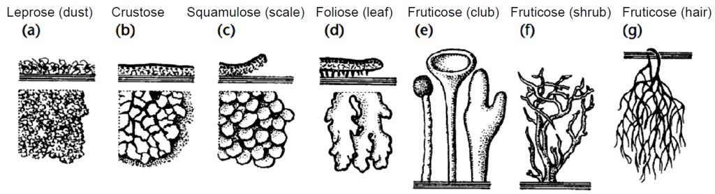
Growth forms
Morphological classification systems that focuses on the shape, substrate adherence and tissue organization of lichens rather than on evolutionary relationships. The simplest system recognizes foliose (leaf-like), fruticose (hair, club or shrub-like), and crustose forms. While imperfect (some lichens inevitabley fall into grey areas), understanding growth form helps you to identify the major tissues, to key out lichens, and to begin to think about their relationship with the environment.
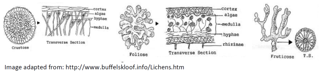
Goward (1999) uses the 7 growth form system illustrated below. Line drawing reproduced courtesy of BC Ministry of Forests and T. Goward, from Goward 1999, fig. 9
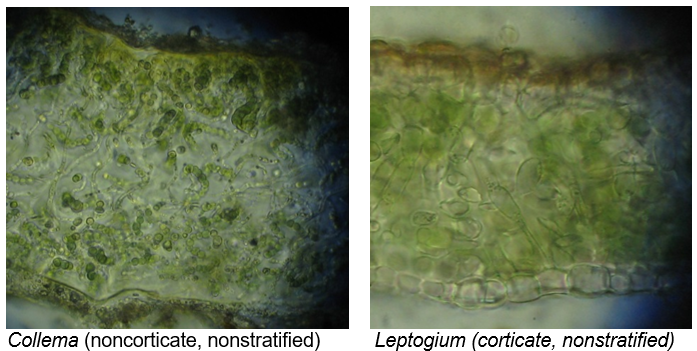
Heteromerious (adjective)
Stratified thallus, where fungal hyphae and photobiont cells are organized in clear layers. Refer to entry for cortices.
Homiomerious (adjective)
Nonstratified thalli, where fungal hyphae and photobiont cells are intermixed rather than in defined layers. May or may not have cortices.
Hypha (plural: hyphae)
Fungal filaments, composing the bulk of the biomass of most lichens.
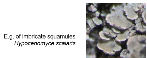
Imbricate (adjective)
Shingled or overlapping.
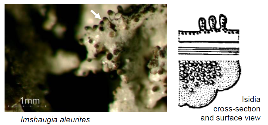
Isidium (plural: isidia)
Asexual reproductive or carbon storage outgrowths typically from the upper surface, composed of both fungal threads and photobiont cells. Isidia typically are wart-like, shiny, and do not detach from the thallus when rubbed, while soredia detach and leave powdery residue on your finger. Line drawing reproduced courtesy of BC Ministry of Forests and T. Goward, from Goward et al. 1994, fig. 9g.
Lichen (plural: lichens)
Symbiotic partnerships between at least one species of fungus and algae. The fungus typically provides the structure or scaffolding, while the alga provides energy in the form of sugar through photosynthesis. These partnerships are incredibly diverse – Alberta is home to over 1,000 species – and they take a wide diversity of forms. Lichens are ecologically important because they provide food for animals, habitat for insects, fix atmospheric nitrogen, stabilize soil, sequester carbon, amongst myriad other roles.
Lobule (plural: lobules)
Little lobes, may function as extra photosynthetic surfaces and/or large vegetative reproductive propagules.
Medulla
The area of (typically) loosely-packed fungal hyphae within a thallus. Refer to entry for cortex.
Mycobiont
The fungal partner(s) of a lichen.
Peltate (adjective)
Attached centrally. Typically used to describe the secondary squamules and how they attach to podetia in some Cladonia species. Peltate squamules may be located inside of the cups (e.g., Cladonia pyxidata and C. pocillum) or on the stalks (Cladonia macrophylla).
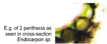
Perithecium (plural: perithecia)
Flask-shaped fungal sexual reproductive structure, often sunken into the thallus and visible only by the opening (=ostiole), which will look like a black pit or dot.
Photobiont
The photosynthetic partner(s) of a lichen.
Placodioid
Intermediate between squamulose and crustose; flattened and radiating out from a center, possibly raising up to being leaf-like at the outer edges but ecorticate under any raised part
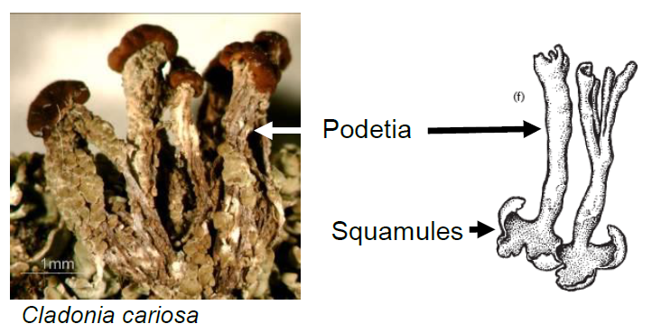
Podetium (plural: podetia)
Erect, stalk-like structures, often bearing cups and/or apothecia, as seen in the genus Cladonia. Typically, podetia arise secondarily from a primary thallus such as squamules or a crust. The primary thallus may persist or disappear after podetia form. Line drawing reproduced courtesy of BC Ministry of Forests and T. Goward, from Goward 1999, fig. 8f.
Pruina (adjective: pruinose)
Powdery or scaly spots on the upper thallus surface, typically made of deposits of dead cells and calcium oxide. Compare to maculae.
Pseudocyphellum (plural: pseudocyphellae)
Small irregular pits in the cortex through which the medulla shows. Pseudocyphellae translates to “false window” and they lack a ‘frame’ or lining of specialized cells found in true cyphellae.
Pycnidium (plural: pycnidia)
Small barrel or flask-shaped growths that contain asexual spores. Can resemble isidia. May be on the surface or embedded within the thallus.
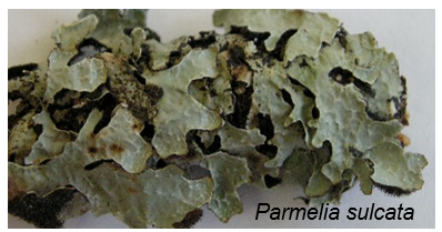
Reticulate
Raised network on surface as seen in Parmelia sulcata or in the pattern of veins on the underside of some Peltigera species such as P. membranacea.
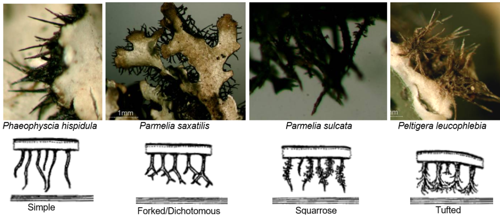
Rhizine (plural: rhizines)
Anchoring hyphae or groups of hyphae, arising from the lower surface. Line drawings of attachment cross-sections reproduced courtesy of BC Ministry of Forests and T. Goward, from Goward et al. 1994, fig. 6a,b,c and e. Rhizines can be very helpful in distinguishing some genera and species (e.g, Peltigera). Adjectives include: squarrose (with perpendicular side branches like a pipe cleaner), thyrisiform (loosely spreading branches that branch compoundly at least two times), dichotomous or forked, or simple (no branches).
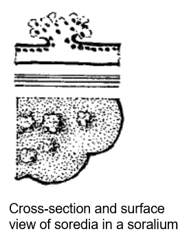
Soralium (plural: soralia)
Patches of powdery or granular propagules known as soredia. The position of soralia on the thallus is a key characteristic.
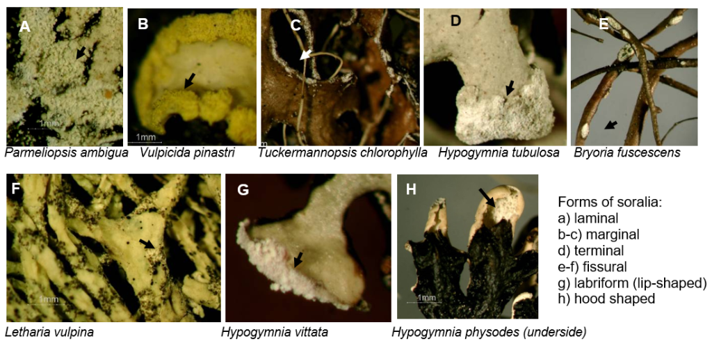
Soredium (plural: soredia)
Small, rounded, asexual reproductive structures made of hyphae and a few photobiont cells. Soredia originate from the algal layer and become exposed through a crack or hole in the cortex. Refer to farinose and soralia. Soredia detach and leave powdery residue on your finger when rubbed. Compare to isidia. Line drawing reproduced courtesy of BC Ministry of Forests and T. Goward, from Goward et al. 1994, fig. 9f.
Squamule (plural: squamules)
Small, flat, leaf-like lobes. In general, squamules are attached at one edge and have a white lower surface, without any attachment structures. Refer to entry for podetia. Squamules may also be peltate (=attached centrally).
Squamulose (adjective, growth form)
Like small foliose lichens but always lacking a lower cortex; may be imbricate (=shingled) or peltate (=attached centrally).
Squarrose
Like a bottle-brush or pipe cleaner (a main axis with secondary branches arising from and perpendicular to the main axis). Refer to rhizines.
Stratified (adjective)
A lichen thallus consisting of organized layers of algal cells and fungal hyphae, typically (from top to bottom) an upper cortex, algal layer, medulla, and lower cortex. Not all lichens have all layers and not all are stratified – some are formed by a more homogeneous mixture of algal and fungal cells (e.g., Collema). Refer to entry for cortices. Synonym: heteromerious. Antonym: homiomerious.
Thallus (plural: thalli)
The lichen body. Thallus is preferred to individual when referring to a lichen body for two reasons. First, a lichen is made of more than one individual organism. Second, in a colony, it is often unclear where one genetically-distinct lichenized organism starts or ends.
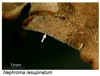
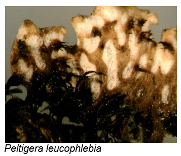
Tomentum (adjective: tomentose)
Erect, cotton-like or spiderweb-like ‘hairs’ that are actually fungal hyphae, varying in thickness, length, and branching. Caused by the outgrowth of surface cells into simple to branched hyphae. Commonly seen on the surface of some Peltigera and Nephroma species. Can resemble fleece fabric!
Tripartite (adjective)
A lichen composed of three dominant partners, a fungus (mycobiont), a green algal photobiont and a cyanobacterial photobiont. The cyanobacterium is often sequestered in structures called cephalodia. Genera with tripartite species include Peltigera, Lobaria, Nephroma, and Solorina. Compare to bipartite.
Veins
Lines of darkened thickened areas, with intervening areas being lighter in colour as seen on the lower surface of species of Peltigera. Often associated with rhizines. Not vascular tissue in the sense of vertebrate arteries and veins, but may have an analogous function in moving resources around and stiffening parts of the thallus like lobes bearing erect apothecia.
Umbilicus (adjective: umbilicate)
A thickened central hold-fast, which from the upper surface, often creates a ‘belly-button’ like dimple.
Images
All photos are by Diane Haughland unless otherwise indicated. Line drawings are credited throughout.
References
Brodo, I. M., S. D. Sharnoff, and S. Sharnoff. 2001. Lichens of North America. Yale University Press, New Haven and London.
Brodo, I. M. 2016. Lichens of North America Updated Keys. Yale University Press, New Haven and London.
Goward, T., B. McCune, and D. Meidinger. 1994. The Lichens of British Columbia Illustrated Keys. Part 1 – Foliose and Squamulose Species. Ministry of Forests Research Program, Province of British Columbia.
Goward, T. 1999. The Lichens of British Columbia Illustrated Keys. Part 2 – Fruticose Species. Ministry of Forests Research Program, Province of British Columbia.
Orange, A., P. W. James, and F. J. White. 2001. Microchemical Methods for the Identification of Lichens. British Lichen Society.
