
Secondary Metabolites
Secondary metabolites are extracellular chemical compounds produced by a species that are not necessary to sustain life, but which may be produced in such abundance and with such specificity that we hypothesize they have an important supportive function.
Those functions include:
- moisture regulation: compounds can be hydrophobic or hydrophilic, and thus regulate where water is absorbed or repelled by the lichen thallus
- light regulation: compounds may absorb, screen or reflect potentially harmful ultraviolet light, helping to maintain internal “greenhouse” conditions for the photobiont and protect DNA from damage
- herbivore deterrence: these compounds deter grazers such as gastropods through toxicity or causing a bad “taste”
- allelopathy: compounds that reduce growth or germination in co-occurring plants and bryophytes
- anti-viral, anti-bacterial, or even anti-fungal protection: compounds that enable a lichen thallus to resist infection, decay and death caused by fast-growing saprophytic or parasitic organisms, over potentially decades of life.
Lichens are famous for the diversity of their secondary metabolites, with over 1,000 compounds known to date. Some of these are also produced by other taxa, but many are unique to lichens. They are a critical part of making the lichen symbiosis successful, especially considering how slow-growing and long-lived lichens typically are. Compare how fast a non-lichenized fungal fruiting body such as a mushroom typically decays or is predated!
We think it is largely the fungi that produce these compounds, which are deposited outside of the hyphae in different parts of the lichen thallus. Recent research has shown that the photobionts, endolichenic fungi, and bacteria from the lichen microbiome also produce important secondary metabolites.
Spot Tests
For over a century lichenologists have used chemical spot tests to help identify (or at least narrow down) the type or class of secondary metabolite in lichens. Spot tests typically involve adding a tiny amount of chemical to a fragment of lichen and looking for specific color changes.
The most common spot tests and their abbreviations are:
K: 10% Potassium hydroxide [KOH]: strong base, reducing agent. Replace every 1-2 months.
C: Bleach aka sodium hypochlorite [NaClO]: reactive with organics and inorganics, oxidizing agent. Use undiluted fresh, unscented, preferably laboratory-quality bleach. Replace weekly.
KC: This test involves a combination of K and C. Saturate the lichen fragment with K, then add a drop of C. Most frequently used to detect a pale yellow pigment, usnic acid.
PD: We use Steiner’s solution, a more stable and less toxic form of Para-phenylenediamine: reactive with strong acids and oxidizing agents. Recipe: 1 g PD, 10 g Sodium sulfite, 100 mL distilled water, 0.5-2 mL dishwashing detergent. PD stains surfaces and skin and should be used with caution. Add together and agitate. Replace every few months or when the solution changes from a light pink to red.
UV: ultraviolet light, either long wave length (365 nm) or short wave length (254 nm). Short wave length provides the clearest fluorescence, but the light is damaging to skin and eyes; never look directly at the light or expose your skin. Dedicated ultraviolet light booths protect you by containing the light, and with UV filtering viewing ports.
Method: take a small piece of lichen and put it on a slide against a white background (i.e., piece of paper). Add a tiny amount of the spot testing chemical and if necessary, push the lichen fragment in to the drop with a dedicated toothpick or pin until it is absorbed. Observe through the dissecting scope. Alternatively, when you’re doing a very precise test like a medullary test on a narrow lobe, keep the lobe attached and do the test in situ; standard advice is to then remove the contaminated lichen fragment to your plate so as to avoid contaminating your packet. We typically do this except for K tests on Physcia specimens – because the atranorin darkens over time, it can be easier to interpret the results later on so we often leave the tested lobe intact. When a number of identifications have been completed, dispose of the slide in a labeled hazardous waste container.
Modified technique for C tests: Bleach, funnily enough, bleaches out the color reaction before it can be observed if applied too liberally, leading to false negatives. It’s best to apply fresh bleach to the exposed medulla in situ, and to try the test at multiple places on the thallus if you initially get negative results, exposing fresh medulla for each test (but avoid pock-marking your thallus, you’ll diminish its appearance for accessioning). Apply the bleach with a pre-soaked toothpick tip or a few hairs of a thinned paintbrush or the tip of a dissecting needle. It’s handy to keep a fragment of something you’ve confirmed is C+ around to test your bleach in case you’re unsure of its reactivity, even straight from the bottle.
What exactly do you test: typically either the cortex or the exposed medulla, ideally in an actively-growing, young part of the thallus where the secondary metabolites tend to be concentrated. If you need to test the medulla, it’s best to ‘flay’ the lichen enough that your drop of chemical won’t also involve the cortex and flood the testing zone with cortical secondary metabolites.
How do you record spot test results?
- If you don’t see any color change with a test, note the test abbreviation followed by a “-“.
- E.g, ,with a negative K test, note the result as “K-“
- If you get a positive color reaction, note the test abbreviation followed by a “+” and a description of the result.
- E.g, with a positive PD test that results in a red color change, note the results as “PD+ red”
- For some lichens it’s important to note where you did your spot test on the lichen thallus
- E.g., “K+ yellow medulla, K- cortex”
- Additional descriptors can include “dingy”, “light”, “deep”, “bright”, “fading or disappearing”, “fast” (typically instant color change), “slow” (indicate time required to get reaction)
TIP: When a lichen goes from dry to wet, even with just water, it typically changes color. The hyphae become more hyaline, and the photobiont more visible. If you’re not sure if you are seeing a pale color reaction to a spot test or a color change due to wetting, perform the spot test alongside a wet fragment for comparison.
TIP: Just because you don’t see a reaction does not mean that the metabolite is absent. Low concentrations of metabolite, old or inactive spot test reagents can result in false negatives.
Discard contaminated slides, paper and lichen fragments in well-labelled repositories for hazardous waste disposal.
Click to download a PDF of Spot Test Color Charts v.2025
Lab Safety
Testing does involve chemicals that are corrosive or could otherwise harm you so review the Material Safety Data Sheets for any chemical prior to use.
Safety questions you should be able to answer:
- what are the hazards of each chemical?
- what do I do if I get the chemical on my skin? in my eye?
- where is the closest eyewash station? first aid kit?
- how do I handle a spill? a fire?
Thin Layer Chromatography
Thin layer chromatography, or TLC for short, is a well-established method to document complex combinations of compounds in lichens with more certainty and subtlety than spot tests allow. Because they require the use of toxic, flammable solvents and peroxide-forming chemicals (which can explode if they crystallize), TLC should only be conducted in a fumehood, in a laboratory, with a strict standard operating procedure and safety plan.
The basic concepts are simple.
- compounds within the lichen are dissolved into a solvent, typically acetone
- that acetone solution is concentrated using heat and/or desiccation, and then spotted (called loading) along the bottom of a TLC plate
- TLC plates are made of a thin stationary phase of silica gel over an inert aluminum or glass backing
- the plate is placed in a shallow layer of a mobile solution system; the solution should not cover your lichen solvent spot.
- the mobile solution wicks up the plate, taking the compounds in the lichen acetone solution with it
- the relative affinity of the lichen compounds to the stationary phase (the silica gel) versus the mobile phase (the solvent system) determine how fast and far the metabolite migrates up the plate relative to the solvent
- typically we do this process simultaneously in 2-3 different solvent systems, each designed to help separate structurally-similar metabolites
- plates are then air dried in a fumehood to remove as much residual solvent as possible
Interpretation is where TLC gets tricky. To characterize each resultant well-defined spot we photograph the plate under various conditions and record:
-
- vertical retention factor: running standardized solutions of reference compounds, allows us to quickly categorize a compound relative to the standard, typically in classes from 1 (hardly moved up the plate) to 7 (moved quite a bit up the plate, with similar retention to the metabolite atranorin
- initial color and fluorescence under ambient light, short wave and long wave light
- opacity: the plate is sprayed with water, many compounds and the stationary phase will be more translucent with fatty acid compounds remaining more opaque
- we then acid-char the plate (brush it with 50% sulfuric acid and slowly heat it on a hot plate or low temperature oven, with good fumigation) and again photograph and record color and fluorescence under ambient light, short wave and long wave light
Using the recorded data, we can query published databases or reference documents to find metabolites that match our observed TLC profile.
Many samples can be examined on a single plate. The concentration of the solvent, and the concentration in the lichen, both can impact TLC results.
Chicita and Bill Culberson (Duke University) were instrumental in developing TLC methods for lichenologists, and documenting the characteristics of known compounds for reference.
-
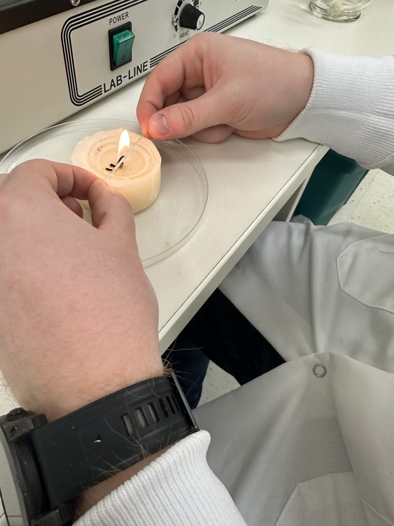
- TLC – pulling capillary tubes for loading lichen metabolites dissolved in acetone onto a TLC plate
-
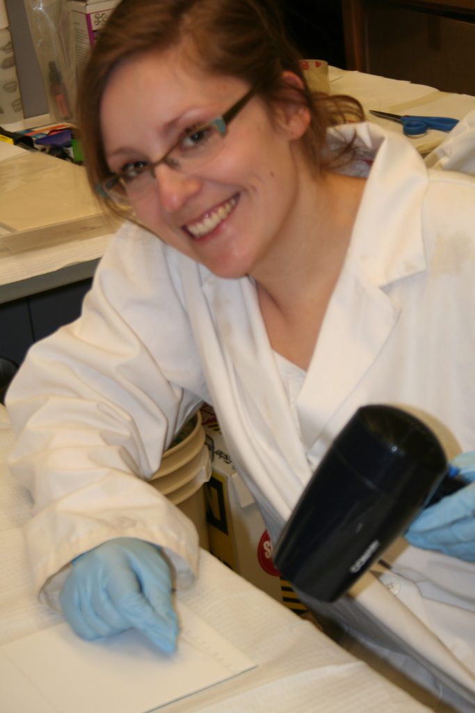
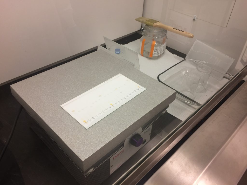
- Darcie loading TLC plate. The hair dryer is one method of creating small, concentrated spots more quickly.
-
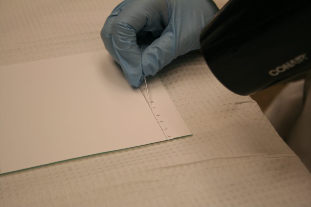
- Spotting or loading a TLC plate
-
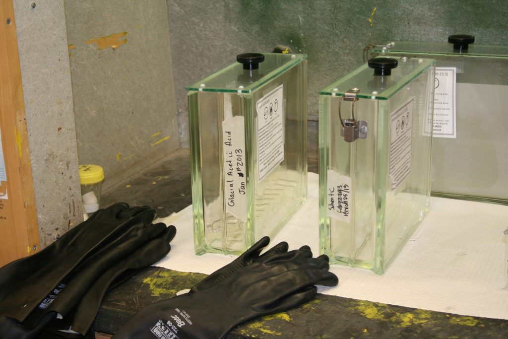
- TLC chambers in a fumehood
-
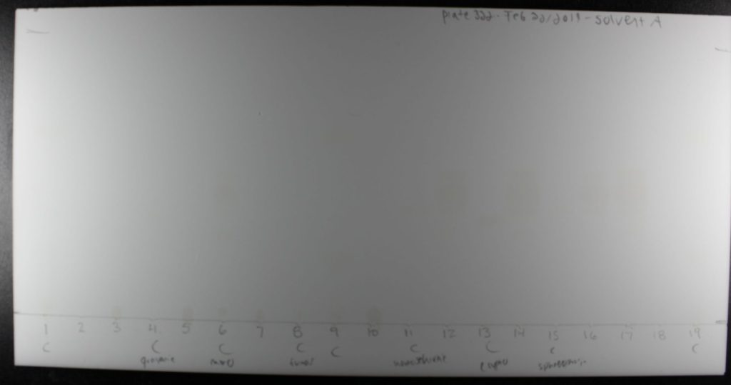
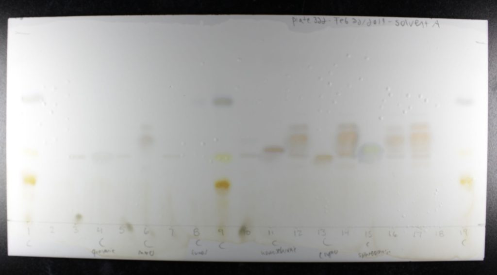
- TLC plate right after running in solvent A. Notice that there is little visible evidence of metabolites at this stage.
-
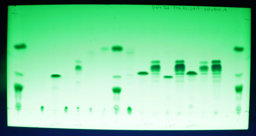
- TLC plate before acid char, in 254 nm light. Each dark spot is a chemical that migrated vertically from a loaded lichen/solvent spot
-
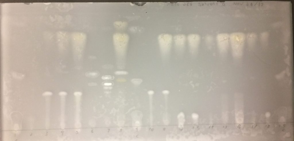
- TLC plate after weting to check for fatty acids
-
- TLC plate starting acid char
-
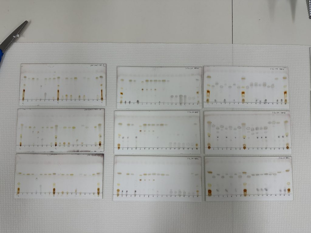
- Completed TLC plates after acid char
-
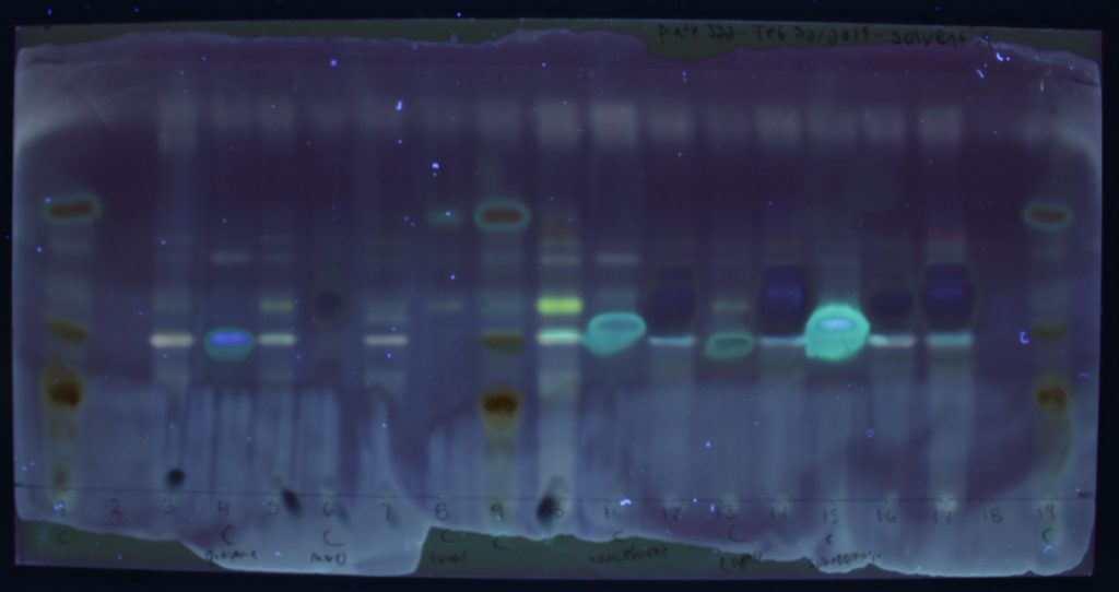
- TLC plate after acid char, in 365 nm light. Note the different fluorescing colors in different spots.
-
- TLC plate after acid char, in 365 nm light. Note the enriched colors!
Instructional Videos
Key Resources
